
The treatment of bone defects has always been a hot topic in clinical practice, and researching different strategies for implantable bone repair materials has been a popular direction. Currently, many researchers are engaged in related studies, continuously exploring innovations to tackle this challenge. 3D printed polycaprolactone (PCL) scaffolds are widely used in bone tissue engineering but suffer from difficulties in cell adhesion, insufficient osteogenic activity, and poor immunomodulatory capabilities. Enhancing the biological responsiveness of PCL scaffolds is a key focus in bone tissue engineering research.The team led by Xu Weikang from the Institute of Biological and Medical Engineering, Guangdong Academy of Sciences, ingeniously prepared composite scaffolds using strontium (Sr) doped bioactive glass (SrBG) with PCL, and modified the surface with polydopamine (PDA) to enhance its osteogenic activity and immunomodulatory capabilities. This opens up new pathways for bone defect repair.

1. Main Content
Ideal bone repair materials must possess biocompatibility and strong osteogenic differentiation capabilities. However, PCL is limited by its insufficient osteogenic ability, high hydrophobicity, and lack of adequate cell adhesion sites. This study prepared a PDA/SrBG/PCL (PSBP) composite scaffold, which was confirmed through in vitro and in vivo experiments to possess multifunctional characteristics of immunomodulation and osteogenic induction.This overcomes the clinical limitations of traditional PCL scaffolds, marking a shift in bone tissue engineering from mere structural replacement to functional reconstruction, providing valuable references for the future development of advanced bone repair materials.
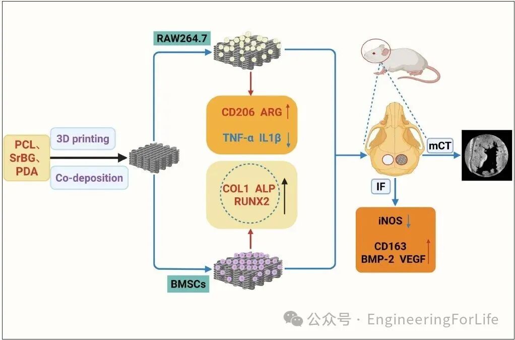
Figure 1. Schematic diagram of the research design
SEM observations of the scaffold’s surface morphology showed that after the addition of SrBG and PDA modification, the PSBP scaffold’s surface adhered to SrBG particles, with smaller and fewer surface pores, resulting in a smoother surface. The average pore size of the PSBP group scaffolds was the smallest. The high porosity of the scaffolds is beneficial for cell adhesion, growth, and vascular ingrowth. The PDA in the PSBP scaffold filled the fiber pores, allowing it to withstand larger mechanical loads, significantly enhancing the compressive strength of the fibers and reducing their degradation rate. The surface modification with PDA improved the scaffold’s hydrophilicity, adhesion, and biocompatibility.
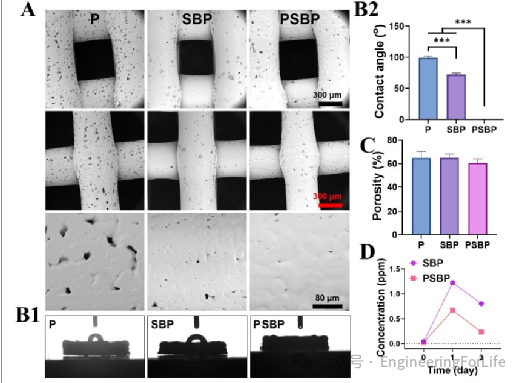
Figure 2. Characteristics of each group of scaffolds. (A) SEM images of the scaffold surface. (B1 and B2) Contact angle and hydrophilicity analysis of the scaffolds. (C) Pore analysis of the scaffolds. (D) Sr²+ release from the scaffolds.
The biocompatibility of the scaffold is a prerequisite for achieving biological functions. In vitro experiments showed that the PSBP scaffold exhibited excellent biocompatibility. Live/dead cell staining results indicated that the fluorescence on the PSBP scaffold was more pronounced than that on the P and SBP scaffolds, with more BMSCs adhering and proliferating on the PSBP scaffold, indicating no cytotoxicity. Meanwhile, CCK-8 proliferation assay results showed that the PSBP scaffold significantly promoted BMSC proliferation compared to other scaffold groups, consistent with the live/dead cell staining results.
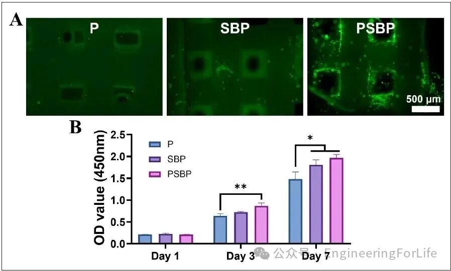
Figure 3. Viability assessment of bone marrow mesenchymal stem cells on the scaffold. (A) Live/dead cell staining of bone marrow mesenchymal stem cells co-cultured with the scaffold for 3 days. (B) Cell proliferation on the scaffold on days 1, 3, and 7.
In terms of anti-inflammatory properties, the PSBP scaffold also demonstrated excellent performance. In macrophage polarization, it significantly promoted the polarization of RAW264.7 cells to the M2 phenotype, which has anti-inflammatory and pro-healing effects, while effectively inhibiting their polarization to the pro-inflammatory M1 phenotype. At the gene expression level, M2 macrophages cultured on the PSBP group showed a significant increase in the expression of cluster of differentiation 206 (CD206) and arginase (ARG) compared to the other two groups. In contrast, the expression of the M1 polarization-related inflammatory genes tumor necrosis factor-alpha (TNF-α) and interleukin-1 beta (IL-1β) was significantly lower in the PSBP group than in the other groups, indicating that the PSBP scaffold can effectively prevent excessive inflammatory responses from macrophages. ELISA experiments showed that the PSBP scaffold more significantly inhibited the expression of the pro-inflammatory factor IL-12 and increased the expression of the anti-inflammatory factor IL-10, suggesting that the relatively low concentration of Sr²+ in the PSBP scaffold is conducive to better coordination of the immune microenvironment.
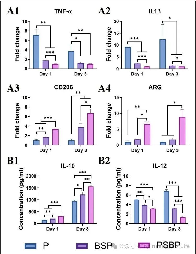
Figure 4. In vitro immunomodulation of related gene expression in M1 and M2 type RAW264.7 cells.
Ideal bone repair materials must possess biocompatibility and strong osteogenic differentiation capabilities. This study incorporated SrBG into the PCL scaffold and modified the scaffold with PDA to detect osteogenic-related genes. From the ALP activity and staining results, it can be seen that the PSBP scaffold strongly induced osteogenic differentiation. Furthermore, qRT-PCR results showed that under osteogenic induction conditions, BMSCs co-cultured with the three scaffold groups exhibited significantly upregulated expression of osteogenic-related genes COL1, ALP, and RUNX2 in the PSBP scaffold group compared to the P and SBP scaffold groups. This observation is consistent with the ALP activity and staining results, indicating that the PSBP scaffold significantly promotes BMSC osteogenic differentiation and mineralized nodule formation.
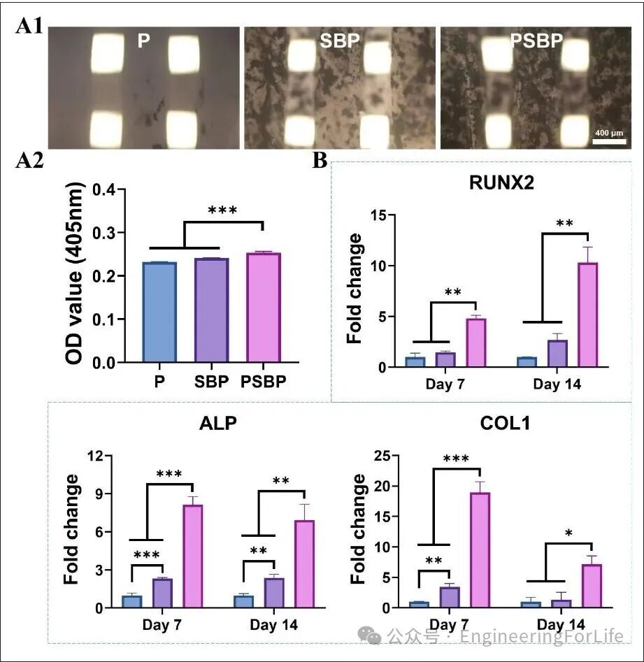
Figure 5. ALP staining (A1) and ALP activity analysis (A2) of the scaffold co-cultured with bone marrow mesenchymal stem cells. (B) Expression of osteogenic-related genes in the scaffold co-cultured with bone marrow mesenchymal stem cells.
In in vivo experiments, researchers established a rat cranial defect model and implanted different scaffolds into the defect sites. To assess the biological safety of the scaffolds, liver and kidney tissues from each group of rats were sampled at 1, 2, and 3 months post-surgery, and tissue sections were stained with HE. Observations revealed that the specific structural morphology and boundaries of the liver and kidneys in each scaffold group were clear, with no signs of inflammatory cell infiltration. This indicates that the composite scaffold has good biocompatibility and no toxic effects.
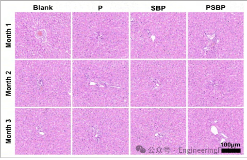
Figure 6. HE staining of liver tissues from rats in each scaffold group.
Micro-CT was used to observe the new bone growth in the bone defect area. Quantitative indicators such as bone mineral density and bone volume fraction were used to evaluate the bone repair effect under a microscope. The 3D reconstruction images showed that the PSBP scaffold, as a bone defect filling material, exhibited significant new bone growth. Similarly, the results of bone volume fraction and bone density analysis were consistent with the Micro-CT results, verifying that the PSBP scaffold has better bone repair effects compared to other scaffolds.
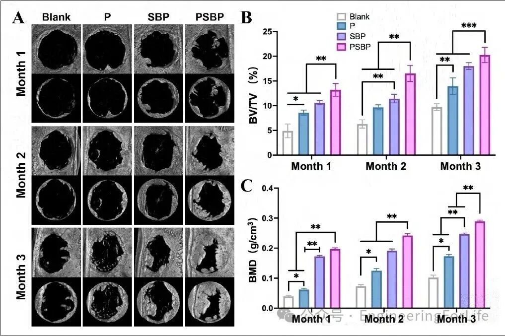
Figure 7. Micro-CT evaluation of bone defect repair effects. (A) Micro-CT analysis of cranial defect areas in each scaffold group. (B) Bone volume fraction (BV/TV%) in the bone defect areas of each scaffold group. (C) Bone mineral density (BMD) in the bone defect areas of each scaffold group. (C) Bone mineral density (BMD) in the bone defect areas of each scaffold group.
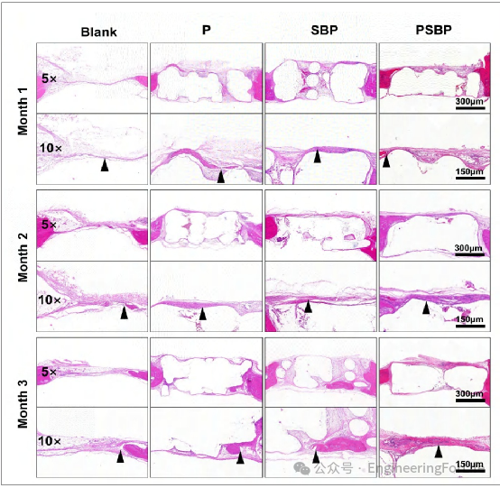
Figure 8. HE staining of bone defect areas in rats from each scaffold group.
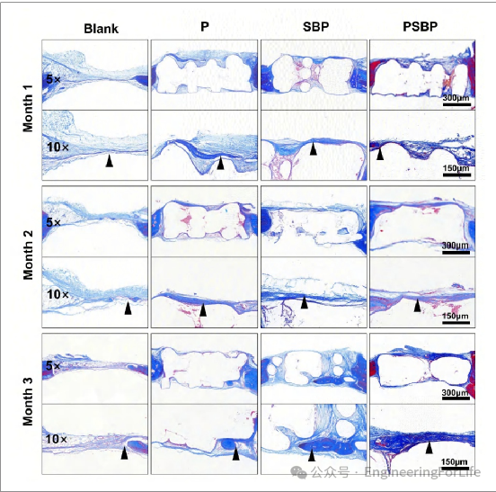
Figure 9. Masson staining of bone defect areas in rats from each scaffold group.
Immunohistochemical staining results also indicated that the PSBP scaffold significantly upregulated the expression of the M2 marker CD163 and downregulated the expression of the M1 marker iNOS, indicating that the PSBP scaffold promoted the polarization from M1 to M2, and the PSBP scaffold exhibited good immunomodulatory properties. Additionally, the PSBP scaffold promoted the expression of BMP-2, suggesting strong osteogenic induction capabilities; it also promoted the expression of VEGF, highlighting its synergistic role in promoting angiogenesis, providing favorable conditions for bone regeneration.
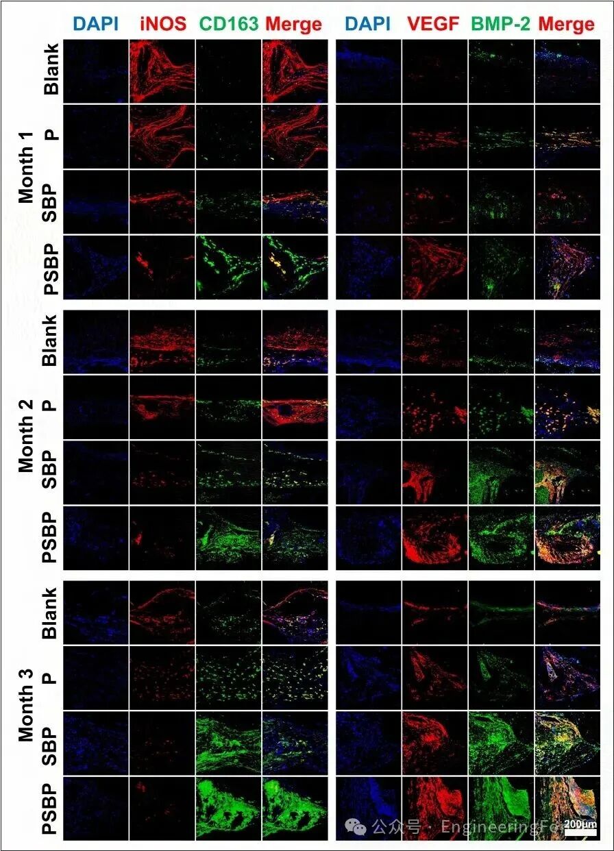
Figure 10. Immunofluorescence staining of iNOS, CD163, VEGF, and BMP-2 in the bone defect areas of rats from each scaffold group.
The composite scaffold prepared in this study can better regulate the immune microenvironment, promote vascular regeneration, and facilitate bone defect repair. Nevertheless, this study has several limitations, including a short observation period and a lack of studies using large animal models in vivo. Further validation is needed to promote its clinical application.
2. Summary and Outlook
This study prepared a PDA/SrBG/PCL composite scaffold with multifunctional characteristics of material modification, immunomodulation, and osteogenic induction, promoting in vitro bone differentiation and bone repair in a cranial defect model in vivo. The composite scaffold is expected to overcome the clinical limitations of traditional PCL scaffolds, marking a transition in bone tissue engineering from mere structural replacement to functional reconstruction, providing valuable references for the future development of advanced bone repair materials.
References:
doi: 10.36922/IJB025210211
Source: EngineeringForLife
Disclaimer: The views expressed are solely those of the author and are for research purposes. The author’s level is limited; if there are any scientific inaccuracies, please leave comments below!