

In the previous issue, we introduced the selection of probes in ultrasound, as well as the impact of dynamic range and grayscale maps on images. In this issue, we will continue to introduce parameters that need to be adjusted according to the organs being examined and areas of interest. Many of these parameters have different names across various manufacturers, but the mechanisms of their effects on images are the same.


Acoustic Energy
By changing the voltage applied to the piezoelectric crystal, the energy of the incident sound wave can be altered. This is the only parameter that can directly change the energy of the sound wave. The greater the acoustic energy, the stronger the reflected sound wave energy, including noise. Therefore, during scanning, it is necessary to minimize acoustic energy while ensuring sound wave penetration.

Total Gain
Total gain adjusts the amplification level of echo signals, directly affecting the overall brightness of the image. When total gain increases, the image becomes brighter overall.
Total gain does not have a fixed set value and is somewhat subjective. The general principle is not to adjust it too high or too low. A gain setting that is too low will cause loss of image details, while a gain that is too high will cause excessive noise that obscures image details, as shown in the figure.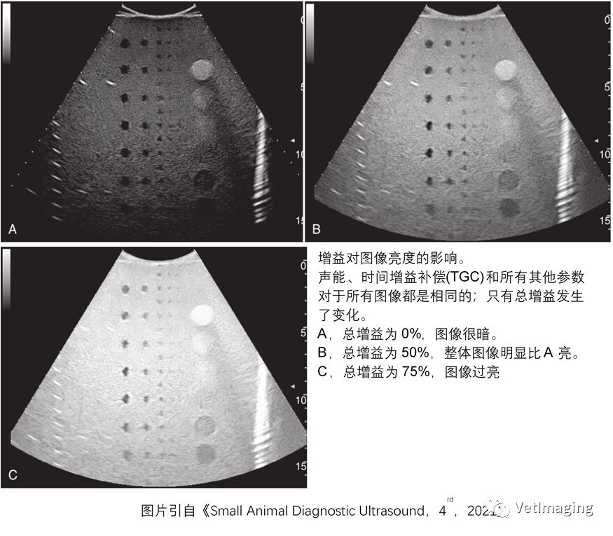

Time Gain Compensation (TGC)
As sound waves propagate, they attenuate with distance, so the echo signals from deeper tissues may be weaker. Time Gain Compensation adjusts the amplification level of echo signals at different depths, compensating for the weakening of signals from deeper tissues due to sound wave attenuation. Proper adjustment of TGC is essential and forms the basis for correct image display. Generally, the time gain compensation should present a smooth curve from left to right.


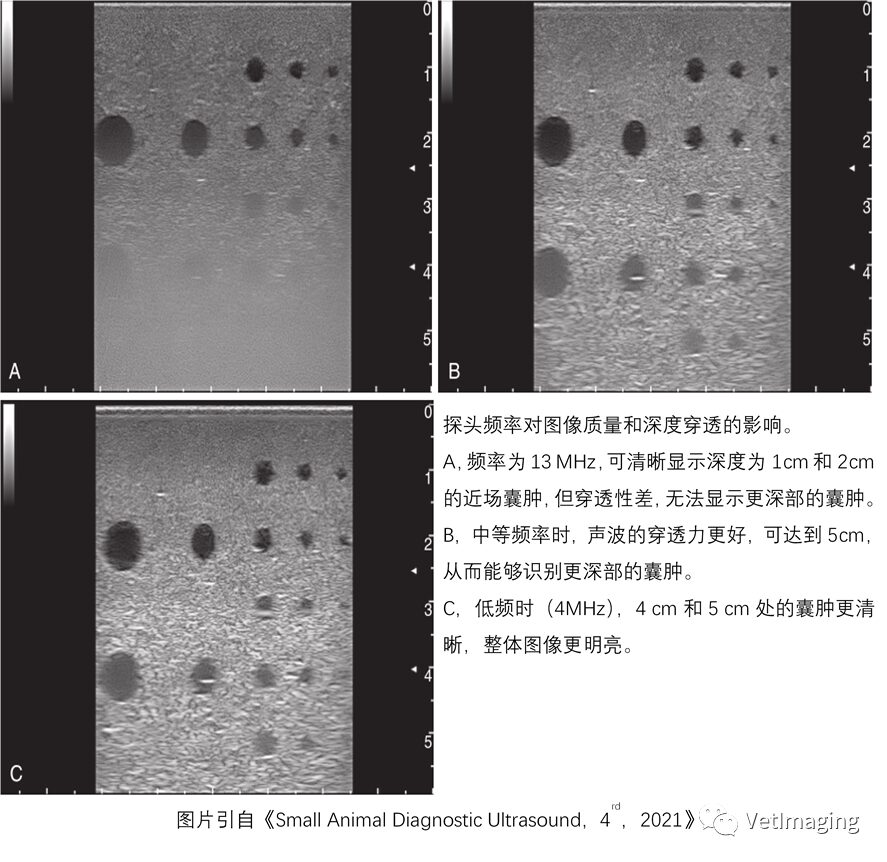
Frequency
The frequency mainly affects the longitudinal resolution of the image. The higher the frequency, the shorter the wavelength, and the higher the longitudinal resolution. However, as the frequency increases, the sound wave’s penetration depth decreases. Therefore, adjust the frequency to the highest level without affecting the strength of far-field echoes.

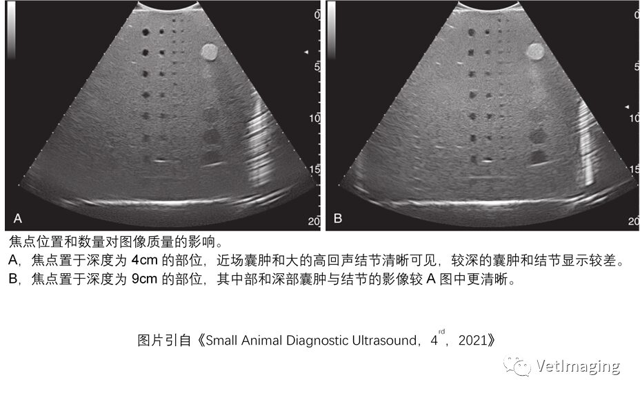
Focus
The focus or focal area is the narrowest part of the sound beam, affecting the lateral resolution of the image. The lateral resolution is best at the focus, so the focal area should always be adjusted to the area of interest or slightly below it.


That concludes our introduction to common parameters for optimizing images. In clinical practice, ultrasound doctors need to select the appropriate probe and make suitable parameter adjustments to optimize the image. Besides the above parameters, many modern ultrasound devices have various special imaging modes to further optimize images. For specifics, please refer to the second issue of the “Ultrasound Diagnosis” topic. In the next issue, we will introduce the Doppler principle.


Zhang Xu
Licensed Veterinarian

If you like it, follow us!