The World Under the Microscope——
Immunocytochemistry
Definition:
Immunocytochemistry (ICC) is a technique established on the basis of immunology, biochemistry, and microscopy technology. It detects the presence and distribution of certain polypeptides, proteins, and large molecular substances such as membrane surface antigens and receptors in cells according to the specific binding characteristics between antigens and antibodies.
Experimental Principle:
ICC is based on the principle of antigen-antibody reaction. A known antigen or antibody is first labeled with a fluorescent group, and then this fluorescent antibody (or antigen) is used as a probe to check for the corresponding antigen (or antibody) in the cells or tissues. Using fluorescence microscopy, one can observe the cells or tissues where the fluorescence is located, thus determining the nature and localization of the antigen or antibody, and using quantitative techniques (such as flow cytometry) to measure the content. It provides a semi-quantitative method to analyze the relative abundance, conformation, and subcellular localization of target antigens. The research samples are usually cultured cell lines, primary cells, or suspended cells obtained from patients and animals, including cell smears, blood smears, swabs, and extracts.
The traditional ICC technique uses chromogenic methods to detect, with enzyme-conjugated antibodies converting chromogenic substrates into colored precipitates at the reaction sites. However, with the advent of immunofluorescence labeling (IF), its application has become more widespread, and in many cases, ICC/IF (Immunocytochemistry/Immunofluorescence) is often used together.
Experimental Steps
01
Cell Preparation: For adherent cells, during passaging, inoculate the cells into culture dishes with pre-treated coverslips. After the cells grow to near confluence, remove the coverslips and wash with PBS twice; for suspension cells, take logarithmically growing cells, wash with PBS by centrifugation (1000rpm, 5min) twice, and prepare cell slides or directly make cell smears.
02
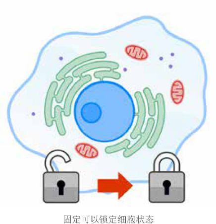
Fixation: Choose an appropriate fixative to fix the cells as needed. The fixed cells can be stored in PBS containing sodium azide at 4°C for 3 months. Wash with PBS 3*5 times.
03
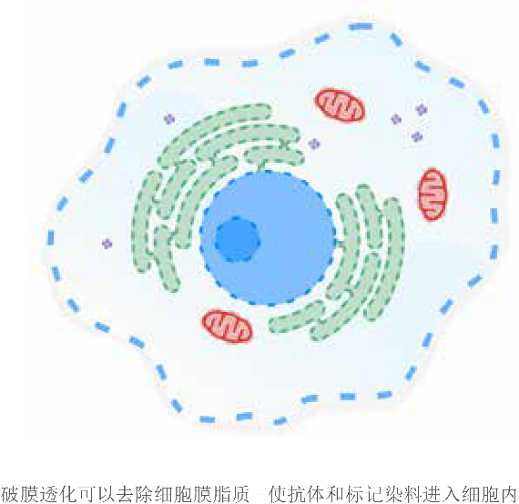
Permeabilization: Cells fixed with crosslinkers (such as formaldehyde) generally need to be permeabilized before antibody incubation to ensure that the antibodies can reach the antigen sites. The choice of permeabilizing agent should fully consider the nature of the antigen protein. The permeabilization time is generally 5-15 minutes. After permeabilization, wash with PBS for 3*5 minutes.
04
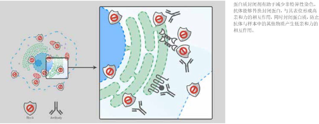
Blocking: Blocking can reduce non-specific binding of antibodies or dyes to the sample. Generally, 2-10% BSA or 5-10% serum is used for blocking, typically for 30 minutes.
05
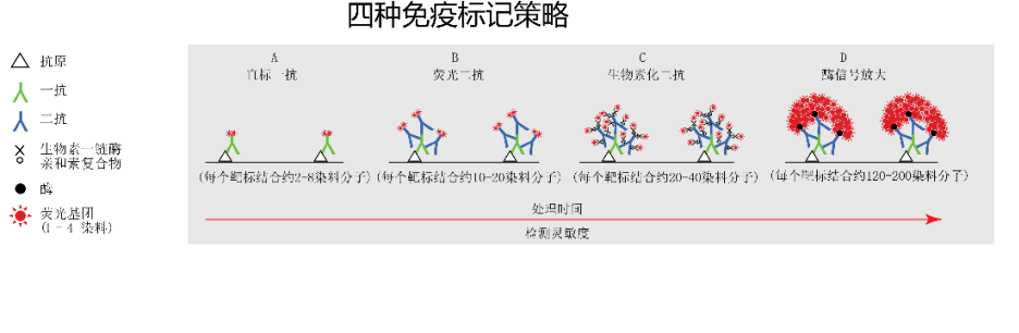
Primary Antibody Binding: Incubate at room temperature for 1 hour or overnight at 4°C. Wash with PBST 3 times, each wash for 5 minutes.
06
Secondary Antibody Binding: Indirect immunofluorescence requires the use of a secondary antibody. Incubate in the dark at room temperature for 1 hour, wash with PBST 3 times, each wash for 5 minutes, then rinse once with distilled water.
07
Mounting and Detection: Add a drop of mounting medium, mount the slide, and check under a fluorescence microscope.
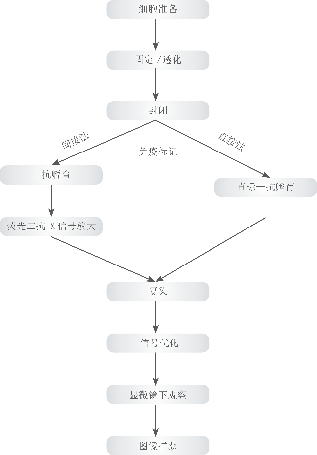
Figure | Experimental Steps
References
Invitrogen ICC/IF workflow guide brochure.
#
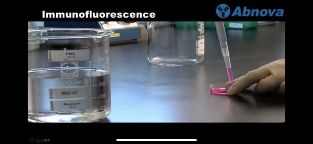
Click the link or read the original text to jump to the learning video!
[Immunofluorescence – Bilibili] https://b23.tv/op4ucSM
/

Text: Scientific Research Branch Three
Huang Ting, Feng Ruiling
Typesetting: Chen Yue
Review: Feng Ruiling, Yao Ranran, Xu Xiaofang
Previous Reviews
Scientific Research Popularization | How to “Seal” Cells Under the Epidemic – Cell Cryopreservation
Scientific Research Popularization | Have You Really Mastered This Basic Skill of Cell Counting?
Scientific Research Popularization | Essential for Research: Have You Learned How to Extract RNA?