In 2020, over 410,000 new cases of liver cancer were reported in China, with more than 390,000 deaths due to liver cancer, bringing the death toll close to the number of new cases. This means that for every 11 newly diagnosed cancer patients, 1 has liver cancer, and almost 1 in 8 patients who die from cancer dies of liver cancer.
Compared to other cancers, the incidence of liver cancer is relatively lower, while the mortality rate is higher. This indicates that the treatment outcomes and prognosis for liver cancer are relatively poor. For liver cancer patients, extending survival time and improving quality of life are urgent issues.
Having understood the macro situation of liver cancer, let’s take a closer look at the microscopic world of liver cancer.
Related immunohistochemical markers for liver tumors CK18/CK8 are commonly referred to as hepatocyte-type cytokeratin. They are expressed in almost 100% of hepatocytes, bile ducts, hepatocellular carcinoma (HCC), intrahepatic cholangiocarcinoma (ICC), and metastatic cancers.CK19/CK7 are known as cholangiocyte-type cytokeratins, expressed in ICC but not in HCC, and can be used for the differentiation between HCC and ICC. They are also expressed in metastatic adenocarcinomas.HepPar-1 is found in normal human hepatocytes and most hepatocellular carcinomas, with the vast majority of hepatocellular carcinomas showing positive expression, and no cross-reactivity with other tumors including gastrointestinal tumors.Alpha-fetoprotein (AFP) is not expressed in normal liver tissue and benign liver lesions, and is almost never expressed in liver metastatic cancers. Its expression rate is 25%-50%, and positive responses are negatively correlated with tumor grade.Arginase-1 (Arg-1) is a highly sensitive hepatocyte marker, with a positivity rate greater than 90% in hepatocellular carcinoma and about 5% in liver metastatic cancers. Its sensitivity is higher than that of AFP and HepPar-1.Glypican 3 (GPC 3) is normally expressed in embryonic liver, kidney, lung tissues, and trophoblast layers of placental tissues, and can be seen in hepatocellular carcinoma, yolk sac tumors, choriocarcinoma, and melanoma, but not expressed in other tissues. Its expression is related to differentiation; the worse the differentiation, the higher the positive expression.Glutamine synthetase (GS) is expressed in hepatocytes at the center of liver lobules but not in periportal hepatocytes or around the portal vein. During the progression of hepatocellular carcinoma, the activity of GS mRNA transcription increases significantly, with GS expression levels gradually rising from atypical hyperplastic nodules → early hepatocellular carcinoma → late hepatocellular carcinoma, enhancing the metastatic ability of cancer cells and serving as an independent prognostic indicator for hepatocellular carcinoma recurrence.Heat shock protein 70 (HSP70) is upregulated in the early stages of hepatocellular carcinoma, with expression levels significantly differing from precancerous lesions and non-cancerous tissues, making it one of the more sensitive indicators for diagnosing early hepatocellular carcinoma.CD34 is not expressed in normal liver sinusoidal endothelial cells, but the abnormal “capillarization” of HCC causes diffuse positivity for CD34 (showing a specific elongated or branched pattern, also known as HCC-type CD34 staining), while staining shows focal distribution in benign liver diseases (such as adenomas, chronic liver diseases, or cirrhosis).Polyclonal CEA (pCEA) shows characteristic tubular staining positivity in both benign and malignant liver tumors, and can also be positive in gastric, colon, lung, pancreatic, or other adenocarcinomas, but positive staining is found in the cell membrane or cytoplasm, not in the tubules.Monoclonal CEA (mCEA) has a positivity rate of 60%-75% in ICC, gastrointestinal tubular adenocarcinoma, and liver metastatic adenocarcinoma, while only <11% positivity is found in HCC, showing positive cytoplasmic staining.S100P is expressed in cholangiocarcinoma and pancreatic ductal carcinoma, but not expressed in normal cholangiocytes (pancreatic duct) or reactive hyperplastic cholangiocytes (pancreatic duct), which helps in determining the benign or malignant nature of cholangiocarcinoma (pancreatic duct) biopsy tissues.MUC1 and MUC5AC are both members of the mucin family; MUC1 can be expressed in fetal liver’s intrahepatic bile ducts but is not expressed in normal adult intrahepatic bile ducts; MUC5AC is not expressed in either fetal or adult normal liver tissues. When both MUC1 and MUC5AC are expressed, cholangiocarcinoma atypical hyperplasia and carcinogenesis should be considered. High expression of both suggests the possibility of cholangiocarcinoma metastasis. The expression rate of MUC1 in ICC is 73.8%, while in HCC it is only 7.8%.
Distinction between benign liver lesions and atypical hyperplastic nodules 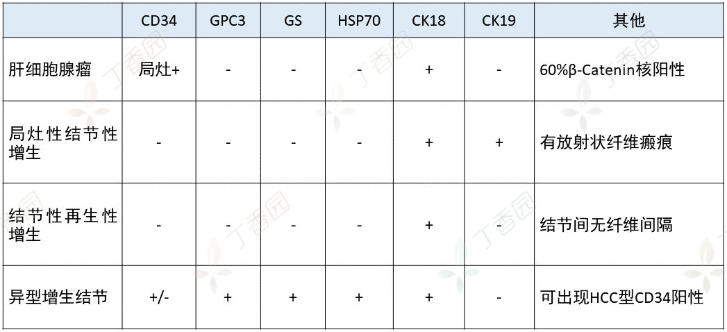 Note: If GPC3 is positive in atypical hyperplastic nodules, especially in highly atypical hyperplastic nodules, it indicates that the nodule is in the late precancerous stage of hepatocellular carcinoma, with a high risk of transformation to hepatocellular carcinoma.
Diagnosis and differentiation of hepatic epithelial tumors Hepatocellular carcinoma (HCC) primarily expresses CK8 and CK18, with very little expression of CK7 and CK19. HepPar-1 is positive in 83.7%; Arg-1 positivity rate exceeds 90%, with sensitivity higher than AFP and HepPar-1; positive for AFP, HSP70, GPC3, pCEA, and GS.Intrahepatic cholangiocarcinoma (ICC) expresses CK7, CK19, CK20, CEA, CDX2, CD15, CA19-9, MUC1, MOC31, and S100P; negative for Arg-1, AFP, and HepPar-1.Mixed hepatocellular and cholangiocarcinoma shows a biphasic immunohistochemical phenotype: HCC area shows positive for Arg-1, AFP, and HepPar-1; while ICC area shows positive for CK7 and CK19. Care should be taken to differentiate from biphasic phenotype HCC (where both hepatocellular carcinoma and cholangiocarcinoma markers are expressed in the same region).Hepatoblastoma shows multidirectional differentiation, expressing various epithelial and mesenchymal cell spectra, negative for HepPar, positive for AFP, GPC3, CK, Vimentin, etc.Neuroendocrine carcinoma expresses CgA, Syn, CD56, etc.
Distinction between primary and metastatic liver tumors Although metastatic liver tumors can originate from any part of the body, the lungs, breasts, colon, pancreas, and stomach are the most common primary sites for metastatic liver tumors.Metastatic tumors usually share common antigen expression with primary tumors. In practical work, a comprehensive analysis can be made by flexibly selecting markers associated with the primary tumor based on the patient’s specific situation, leading to a diagnosis.
 Differentiation between intrahepatic cholangiocarcinoma and metastatic pancreatic ductal adenocarcinoma In liver biopsy specimens, distinguishing between intrahepatic cholangiocarcinoma (ICC) and metastatic pancreatic ductal adenocarcinoma (PDA) is histologically challenging, but differentiation between the two is clinically significant.Is there a simple and easy method for differentiation?A study once selected a group of antibodies including S100P, MUC5AC, Maspin, CK17, IMP3, and pVHL to distinguish between the two, achieving promising results.The results showed:IMP3 and Maspin were highly expressed in both ICC and PDA, lacking diagnostic value;when S100P−/pVHL+/MUC5AC−/CK17− is present, it suggests ICC;when S100P+/pVHL−/MUC5AC+/CK17+ or S100P+/pVHL−/MUC5AC−/CK17+ is present, it suggests PDA.Note: von Hippel–Lindau protein (pVHL): pVHL is a tumor suppressor factor, and its mutation can lead to tumors in various organs or systems, such as the central nervous system, endocrine system, and kidneys.pVHL is a subunit of a multi-protein ubiquitin ligase, which negatively regulates the expression of many hypoxia-inducible genes controlled by hypoxia-inducible factor (HIF). Under physiological oxygen concentrations, pVHL can guide the rapid ubiquitination and degradation of HIF through the ubiquitin-proteasome pathway, preventing inappropriate expression of these hypoxia-inducible genes.Planned by: GoEunCover image: Zcool