ZHEJIANG GONGSHANG UNIVERSITY

LABORATORY of FOOD ORAL PROCESSING,
ZHEJIANG GONGSHANG UNIVERSITY
Food Oral Processing Laboratory (Hangzhou, China)

December 07, 2022
Sensory Physics 101 | Visual Perception or Vision
Introduction
Humans possess five basic senses, including vision, hearing, touch, smell, and taste, to help perceive the surrounding world. The associated body parts transmit sensations to the brain, which then transforms these sensations into information that can be understood by the human body through a complex and mysterious process. Bright colors, loud sounds, intense pain, familiar tastes on the tongue, or fragrant smells—all stimuli converge to create a picture that helps us recognize our environment. The sensory system of the human body is very complex and mysterious, and animals are no exception: felines are known for their night vision, elephants have the most sensitive sense of smell among animals, and bats rely on sound waves for hunting. Magnetoreception—the ability to detect the Earth’s magnetic field—is even considered a sixth sense that some birds, as well as certain mammals, reptiles, and fish, are believed to possess.
The Sensory Physics 101 series aims to explore the physical processes that constitute each sense (including the so-called sixth sense) and explain the extent to which they endow certain species with “superpowers”.
Although recent research from York University suggests that there is no universal hierarchy among the senses[1], vision is often regarded as the most precious sense in contemporary Western culture[2]. In a survey conducted by the Department of Psychology at the University of Regensburg regarding the dominance of vision in research, 75% of participants indicated that they feared losing their vision more than losing their hearing or touch[2]. Bendong[3] defines visual perception as “the ability to interpret the surrounding environment by processing the information contained in visible light.” This process is managed by a highly specialized ocular organ system, and the study of light has evolved into an important branch of physics—optics. Furthermore, controversially, the processing of visual information seems to dominate other forms of information processing[2]. Therefore, this article will begin by describing various physiological structures related to vision and explore the astonishing abilities of cats, barn owls, and mantis shrimp, such as near-night vision[4] and yaw gaze stabilization[5].

Figure 1 Five Senses
First, the first element of the visual system is the eye—a receptor capable of receiving information from distant objects[6]. It is a simple optical instrument, approximately 24 mm in diameter in adults, covered by a flexible tissue called the sclera, and consists of two positive lenses: the cornea and the crystalline lens, which project images onto the retina[7]. The transparent cornea allows light to enter the eye, shaped like a crescent lens, which has two spherical surfaces, one convex and the other concave. To date, the inner surface of the cornea (the interface between air and the eye) has the highest refractive power in the human eye, approximately 48 diopters[8], while a layer of aqueous tear film on the cornea ensures the smoothness of the first optical surface for optimal quality[9].
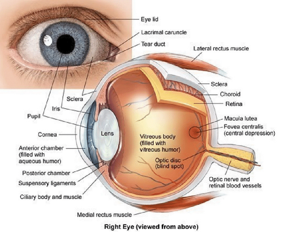
Figure 2 Detailed Anatomy of the Eye
Then, light or photons pass through the black circular opening in the center of the iris—the pupil, which changes size according to the ambient light, from less than 2 mm in bright conditions to more than 8 mm in darkness[9]. Thus, the iris, which has two sets of muscles, acts as a diaphragm, and the entrance pupil (the image of the iris through the cornea) and exit pupil (the image of the aperture through the lens) play a crucial role in imaging and visual quality[8]. Light illuminates the internal lens of the eye, known as the crystalline lens, which combines with the cornea to focus light onto the retina. The crystalline lens is an active optical element that can change its shape to alter the light intensity throughout the eye, allowing the eye to focus on objects at different distances[9].
After passing through the transparent vitreous humor of the eye, light finally reaches the retina, which is the light-sensitive neural tissue on the inner wall of the back of the eye, acting as a screen for image formation[9]. The retina plays a dual role in the optical design of the eye[8]; its curvature closely matches the curvature of the image, thus maintaining reasonable peripheral image quality, and each retinal cone from the mosaic of photoreceptors acts as an individual waveguide, guiding light from the inside to the outside containing visual pigments[10]. Additionally, since photoreceptors near the axis tend to be more efficient than those at oblique angles, they respond more strongly to light entering near the center of the pupil compared to light entering the edges of the eye—this phenomenon is known as the first Stiles-Crawford effect[11]. This reduced sensitivity to peripheral light diminishes their aberrations, which are the differences between the perfect (spherical) and actual wavefronts, leading to blurred or distorted images formed by the lens[7]. Therefore, the retina plays a crucial role in optimizing the performance of the eye’s optical system.
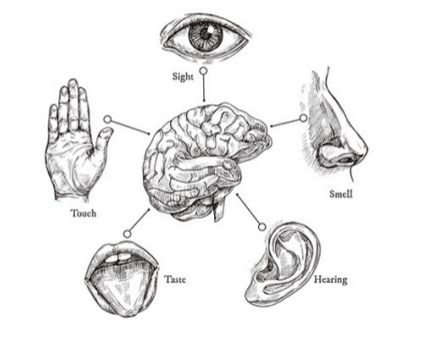
Figure 3 Sketch of the Five Senses in Humans
Thus, while the peripheral part of the retina exhibits lower resolution when detecting motion and objects, the fovea, the center of the visual field, is densely packed with photoreceptors to provide the highest possible resolution[7]. Humans have three types of photoreceptors: intrinsically photosensitive retinal ganglion cells (ipRGCs); rods—responsible for dark vision at low light levels; and cones—providing photopic vision under higher light levels, as well as color vision and high spatial acuity[12]. In the conditions of a 21st-century metropolis, nearly all of our vision is mediated by cone cells, yet cones only account for 5% of the retinal photoreceptors. On the other hand, rods contribute to human vision only under quite limited conditions, such as after prolonged exposure (usually tens of minutes) to very low light levels (dusk or even lower light levels)[12].
The fovea, which is devoid of rods and contains only 100,000 cone cells (0.1% of the total photoreceptors), enables humans to have high visual acuity. Additionally, the cone system can quickly respond to changes in light intensity at dusk and above[12]. The human eye has three types of cone cells: long-wavelength, medium-wavelength, and short-wavelength, peaking at red, green, and blue wavelengths, respectively. At medium and high brightness, these cone cells form the basis of color perception, while under very dim light, monochromatic rod cells dominate[13]. Despite the latter’s poorer performance, they are present in vast numbers on the retina, a phenomenon caused by the primitive rod cells developing the ability to respond reliably to single photons. This forces the retina to process these quantum signals as discrete signals rather than analog signals, thus demonstrating a significant advantage in lowering the visual threshold to extremely low levels[12].
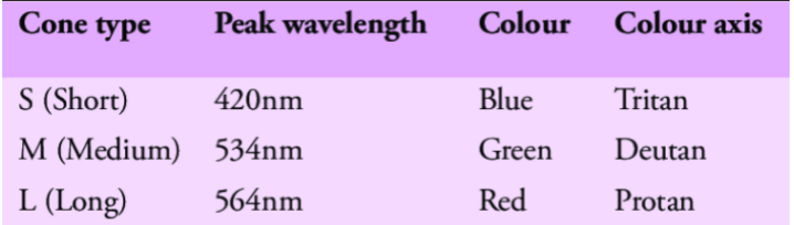
Figure 4 Three Types of Cone Cells in the Human Retina
Finally, photons are converted into electrical impulses by the visual transduction in rods, cones, and intrinsically photosensitive retinal ganglion cells, which are then analyzed by the rest of the visual system.
Intermediate neurons in the retina, such as horizontal cells, bipolar cells, and amacrine cells, transmit information to output neurons—the retinal ganglion cells, of which there are about 30 types, each of which generates action potentials based on the quality and location of visual stimuli in the environment. Thus, action potentials are transmitted via the optic nerve to the brain, where they are transformed into perception and light-mediated behaviors[14]. The brain continues to filter images based on color, contrast, position, and direction of motion, then compares them with memories to interpret what is seen. While humans can quickly recognize objects that vary greatly, this ability is not yet fully understood[15], but increasing evidence suggests that the brain solves such problems through a series of reflexive, feedforward computations, ultimately forming robust neuronal representations in the inferior temporal cortex[16].
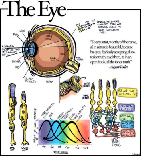
Figure 5 The Eye
While the human visual system is designed very well, some animals have completely different perceptual abilities. For example, felines have improved vision due to distortions in some key components, as mentioned in a 2010 study[4] that aimed to create an educational game simulating human and cat vision in a puzzle game environment—Catalyst. First, in terms of color, cats have only two types of cone cells; although a third type has been discovered, its low quantity does not have a significant impact, meaning that humans can perceive colors and light better than cats. Additionally, the visual acuity that characterizes object discrimination is also weaker in cats, but they have a wider field of view, allowing them to better perceive motion around them. However, the tapetum lucidum (a layer of tissue behind the retina) reflects light and gives cats their phosphorescent eyes, combined with a larger number of rod cell sensors, making the minimum light threshold for cats seven times lower than that of humans. Furthermore, a study from the University of California[17] indicated that the area change of a cat’s vertical slit pupil ranges from 135 to 300 times, while the area change of a human’s round pupil is about 15 times. Banks[17] explains: “Species that are active during the day and night need to fully expand in dim conditions while contracting during the day to prevent glare.” Therefore, while cats cannot see in complete darkness, their eyes are adapted to see in a broader range of light.
The barn owl is also an impressive nocturnal predator, as a 2012 study indicated that the large, forward-facing eyes of the barn owl are the basis of its stereoscopic vision[18]. Additionally, despite significant differences in neural organization compared to humans, the level of visual perception in barn owls is surprisingly similar to that of humans. Barn owls possess superior optical quality and a retina dominated by rod cells, but their visual acuity is reduced to better utilize dim environments for effective predation[18].

Figure 6 A Cat with Goggles
However, the award for the most complex visual system in the animal kingdom should go to the mantis shrimp. Recent experimental research has shown that mantis shrimp have 12 color vision channels, three times more than humans and six times more than cats[5]. Additionally, an earlier paper found[19] that mantis shrimp can perceive polarized light—light waves that vibrate in only one direction, which is not perceived by humans but helps shrimp distinguish prey that blends into the background. Their rotating eye movements also enhance this ability to perceive dynamic polarized light, a capability that has yet to be found in other species.
To further study visual mechanisms, researchers installed a black-and-white striped system along a drum[5], which rotated around an aquarium containing crustaceans. The drum could pitch up and down, twist around a fixed point of focus in front of the eyes, and rotate horizontally, with angles and degrees of rotation far exceeding those of most mammals and insects. The basis of this research is that all animals must maintain gaze stability to accurately perceive motion and depth (using disparity, the difference in images between the two eyes, to indicate near and far objects). The collected data indicated that the eyes of mantis shrimp, while rotating, also performed yaw movements supported by the rotational motion system. This suggests that the rotational motion acts as a gaze stabilizer for yaw rotation, allowing mantis shrimp to continuously rotate their eyes to adjust their lateral view. Therefore, researchers speculate that the nervous system of mantis shrimp can negate the effects of rotational motion to perceive stable images, a goal that both humans and current machines are striving to achieve, making this crustacean the only recorded case of independent visual capability in rotational motion in the animal kingdom, although this feature still lacks a clear rationale for its existence. Furthermore, the study proposed a hypothesis that the visual system of crustaceans is equipped with a radial motion detection array capable of tracking stimuli from directions outside of yaw and pitch settings, which would be an exciting capability for future research.
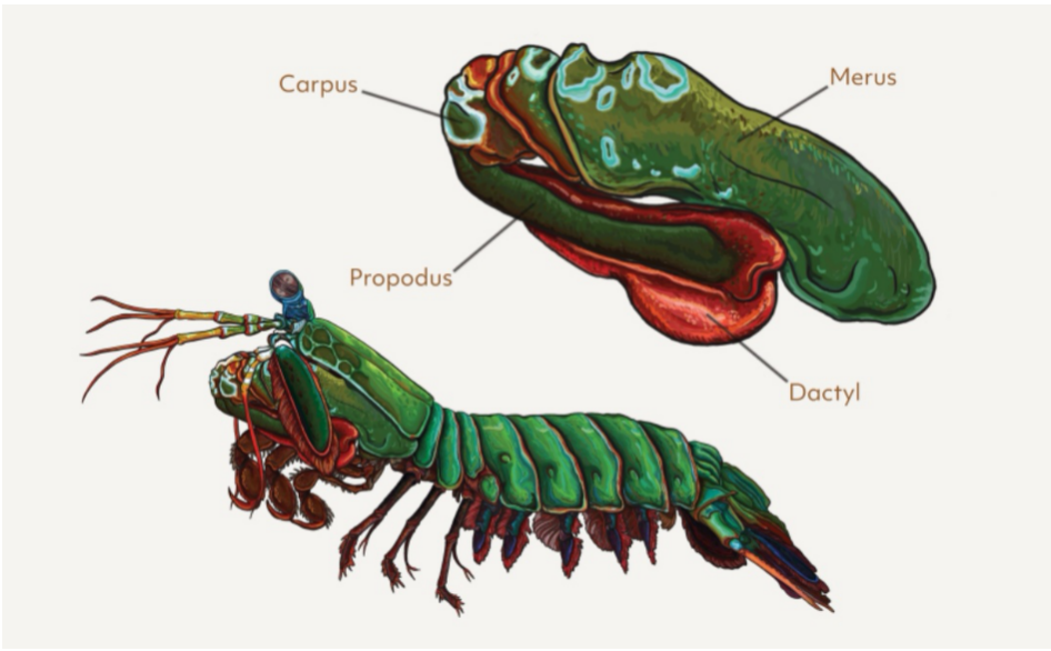
Figure 7 Mantis Shrimp’s Appendages for Prey Capture
In summary, the biophysical system behind vision is a highly complex system, encompassing a series of processes from light refraction in the eye, image projection onto retinal tissue, phototransduction in rods, cones, and ganglion cells, action potential transmission through the optic nerve, to the final complex feedforward computations in the brain to reconstruct comprehensible visual information. The synergy of experimental methods and advanced modeling allows us to gain deeper insights into the connections between optical mechanics, optical design, neurophysiology, and evolutionary demands. In fact, as human eyes evolved towards better night vision, species like cats and barn owls have developed acute nocturnal vision and motion detection abilities to adapt to their predatory lifestyles. However, as exemplified by the mantis shrimp’s incredible visual system, some mysteries of nature remain unsolved. Thus, future research may yield surprising discoveries, potentially leading to breakthroughs in fields such as ophthalmic optics and even neurosurgery.
References
[1]Majid, A., Roberts, S., Cilissen, L., Emmorey, K., Nicodemus, B., & O’Grady, L. et al. (2018).Differential coding of perception in the world’s languages. Proceedings Of The National Academy Of Sciences, 115 (45), 11369-11376. https://doi.org/10.1073/pnas.1720419115.
[2]Hutmacher, F. (2019). Why Is There So Much More Research on Vision Than on Any Other Sensory Modality?. Frontiers In Psychology, 10. https://doi.org/10.3389/fpsyg.2019.02246.
[3]Bendong, L. (2015). The Science of Sense. Department Of Mathematics, Tongji University, Shanghai 200092, China. https://doi.org/DOI:10.13140/RG.2.1.3053.2004.
[4]Long, J., Estey, A., Bartle, D., Olsen, S., & Gooch, A. (2010). Catalyst. Proceedings Of The Fifth International Conference On The Foundations Of Digital Games – FDG ’10.https://doi.org/10.1145/1822348.1822364.
[5]Daly, I., How, M., Partridge, J., & Roberts, N. (2018). Complex gaze stabilization in mantis shrimp. Proceedings Of The Royal Society B: Biological Sciences, 285. https://doi.org/10.1098/rspb.2018.0594.
[6]Sybesma, C. (1989). Biophysics of the sensory systems. Biophysics, 255-279. https://doi.org/10.1007/978-94-009-2239-6_11.
[7]Artal, P. (2016). The Eye as an Optical Instrument. Optics In Our Time, 285-297. https://doi.org/10.1007/978-3-319-31903-2_12.
[8]Navarro, R. (2009). The Optical Design of the Human Eye: a Critical Review. Journal Of Optometry, 2 (1), 3-18. https://doi.org/10.3921/joptom.2009.3.
[9] Artal, P. (2014). Optics of the eye and its impact in vision: a tutorial. Advances In Optics And Photonics, 6 (3), 340. https://doi.org/10.1364/aop.6.000340.
[10] Rativa, D., Vohnsen, B., & Blendowske, R. (2011). Simulating human photoreceptor optics using a liquid-filled photonic crystal fiber. Investigative Ophthalmology & Visual Science, 52 (3206).https://doi.org/10.1364/boe.2.000543.
[11] Nilagiri, V., Suheimat, M., Lambert, A., Turpin, A., Vohnsen, B., & Atchison, D. (2021). Subjective measurement of the Stiles-Crawford effect with different field sizes. Biomedical Optics Express, 12(8), 4969. https://doi.org/10.1364/boe.427834.
[12] Lamb, T. (2015). Why rods and cones?. Eye, 30 (2), 179-185. https://doi.org/10.1038/eye.2015.236.
[13] Davies, A., Morris, D., Kalson, N., Wright, A., Imray, C., & Hogg, C. (2011). Changes to Colour Vision on Exposure to High Altitude. Journal Of The Royal Army Medical Corps, 157 (1), 107-109. https://doi.org/10.1136/jramc-157-01-17.
[14] Laha, B., Stafford, B., & Huberman, A. (2017). Regenerating optic pathways from the eye to the brain. Science, 356 (6342), 1031-1034. https://doi.org/10.1126/science.aal5060.
[15] Rajaei, K., Mohsenzadeh, Y., Ebrahimpour, R., & Khaligh-Razavi, S. (2019). Beyond core object recognition: Recurrent processes account for object recognition under occlusion. PLOS Computational Biology, 15 (5). https://doi.org/10.1371/journal.pcbi.1007001.
[16] DiCarlo, J., Zoccolan, D., & Rust, N. (2012). How Does the Brain Solve Visual Object Recognition?.Neuron, 73 (3), 415-434. https://doi.org/10.1016/j.neuron.2012.01.010.
[17] Banks, M., Sprague, W., Schmoll, J., Parnell, J., & Love, G. (2015). Why do animal eyes have pupils of different shapes?. Science Advances, 1 (7). https://doi.org/10.1126/sciadv.1500391.
[18] Orlowski, J., Harmening, W., & Wagner, H. (2012). Night vision in barn owls: Visual acuity and contrast sensitivity under dark adaptation. Journal Of Vision, 12 (13), 4-4. https://doi.org/10.1167/12.13.4.
[19] Daly, I., How, M., Partridge, J., Temple, S., Marshall, N., Cronin, T., & Roberts, N. (2016). Dynamic polarization vision in mantis shrimps. Nature Communications, 7 (1).https://doi.org/10.1038/ncomms12140.
Click to read the original text and view the original references
”
□ Text Editor:
□ Text Review:
□ WeChat Formatting:
Chen Lele
Zhu Xiaoxuan
Wei Yilu
Professor Chen Jianshe
Email:
Office Address:
Phone:
Fax:
Website:
Laboratory Address:
Zhejiang Gongshang University, College of Food and Biological Engineering, Room 415
(00)86 571 28008904
(00)86 571 28008900
http://spxy.zjgsu.edu.cn/List2.asp?nid=89
Food Oral Processing Laboratory, Zhejiang Gongshang University, 18 Xuezheng Street, Jianggan District, Hangzhou, China, Rooms 337 & 425