What is Functional Magnetic Resonance Imaging?
Various magnetic resonance imaging techniques based primarily on conventional T1WI and T2WI mainly display the morphological structures of human organs or tissues and their signal intensity changes, collectively referred to as conventional MRI examinations or conventional MR imaging sequences. With the development of MRI system hardware and software, various ultra-fast imaging sequences (such as EPI technology) have emerged, reducing the data acquisition time to milliseconds.
MRI examinations based on ultra-fast imaging sequences can evaluate the functional state of organs, revealing physiological information within the organism, collectively referred to as functional magnetic resonance imaging or functional imaging techniques.
These techniques include diffusion weighted imaging (DWI), perfusion weighted imaging (PWI), functional MRI (fMRI), real-time imaging of cardiac motion and perfusion, magnetic resonance spectroscopy (MRS), whole-body imaging, and magnetic resonance microscopy.
What is the Role of the b Value in Diffusion Weighted Imaging?
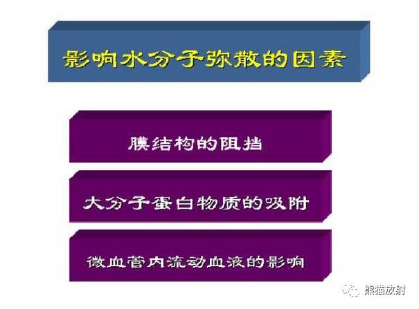
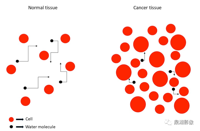
Diffusion is a term that describes the random thermal motion (Brownian motion) of water and other small molecules.
On a macroscopic level, the net movement of water molecules can be described by the Apparent Diffusion Coefficient (ADC), measured by applying two gradient pulses, with an imaging mechanism similar to phase contrast MRA.
The signal intensity changes in DWI depend on the ADC state of the tissue and the strength of the Motion-Sensitive Gradient (MPG). MPG is controlled by the b value (i.e., diffusion gradient factor, also known as b value).
The b value actually determines the share of the ADC contributing to the image contrast, that is, the degree of diffusion weighting.
In the DWI scanning sequence, if a long TR and long TE are used, and b=0, a normal T2WI contrast (SE-EPI) or T2*WI contrast (GRE-EPI) image will be formed. As the b value increases (usually 500~1000 s/mm²), the image contrast gradually shifts from T2 weighting to diffusion weighting.
When the high signal of a lesion in MR images is not due to prolonged T2 time but reflects a decrease in ADC, it forms what is known as DWI. The activation of MPG is the distinctive point between DWI and conventional MRI.
How to Analyze DWI and ADC Images?
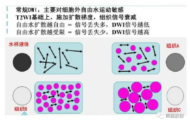
The diffusion weighted sequence scan produces two types of images, namely diffusion images (DWI) and ADC images. In the diffusion image, the signal intensity of the lesion or damaged tissue is often higher than that of normal tissue, while the signal intensity in the area with the highest diffusion freedom is the lowest, making the signal performance of the lesion tissue in DWI resemble that of conventional “T2WI”.
Through post-processing operations on DWI data at the workstation, a grayscale ADC image can be formed. Based on the size of the ADC value corresponding to the signal intensity, a grayscale ADC image is formed. The damaged tissue shows restricted diffusion, with a decreased ADC, appearing as a darker area; the ADC in freely diffusing areas is higher, with relatively bright signal intensity. In the pseudocolor ADC image, when the ADC value decreases, it appears green, normal areas appear orange-yellow or red, and areas with limited water molecule diffusion appear gray.
The ADC image can distinguish whether the high signal displayed by DWI is due to restricted diffusion or due to the tissue having a very long T2 decay time (T2 transmission effect).
What is the Clinical Significance of Diffusion Weighted Imaging?
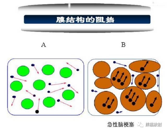
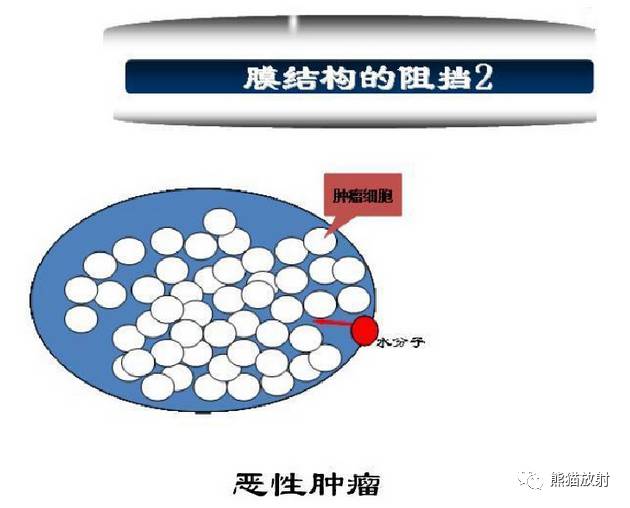
The most valuable clinical application of DWI is in detecting and evaluating acute cerebral infarction and malignant tumors.
In the early stages of cerebral infarction, brain cells swell and absorb water from the extracellular space, restricting the diffusion of water molecules inside and outside the cells, leading to a decrease in the local tissue’s ADC value, with DWI showing a high signal, while at this time, conventional MRI may appear normal. The timing of DWI examination is crucial, as it mainly shows acute lesions within a few days after cerebral infarction.
The ADC value of human tissues generally ranges from 0.2 to 2.9 × 10⁻³ mm2/s. Since the ADC value changes under different pathological conditions, DWI can not only distinguish between acute cerebral infarction with restricted diffusion and non-restricted edema lesions (such as vasogenic edema: venous sinus thrombosis, reversible posterior leukoencephalopathy, etc.) in the brain, but is also commonly used to differentiate between benign and malignant tumors in other body regions.
-
In breast imaging, to differentiate breast cancer, fibroadenoma, and glandular hyperplasia;
-
In liver imaging, to differentiate hepatocellular carcinoma, metastatic tumors, and hemangiomas;
-
In pancreas imaging, to distinguish mucinous tumors from other tumors;
-
In pelvic imaging, to show prostate cancer, endometrial cancer, and cervical cancer;
-
To detect local or distant metastatic lymph nodes;
-
To differentiate the nature of fractures as pathological or traumatic;
-
To evaluate bone contusions;
-
After radiotherapy or chemotherapy for malignant tumors, when DWI shows a decrease in tumor signal intensity or an increase in ADC value, it indicates that the tumor tissue is responding to treatment, suggesting that the treatment is effective.
During signal acquisition in the DWI sequence, if a motion-sensitive gradient is applied in a specific direction, it can form diffusion tensor imaging (DTI). Currently, DWI is widely used to display different orientations of white matter fiber bundles in living brain and spinal cord neural tissues, studying some pathological processes related to white matter lesions.
DWI examinations have certain requirements for the hardware and software configuration of the MRI system, such as mid to high field strength MRI systems, higher gradient switching rates, and good EPI performance. DWI is easily affected by magnetic susceptibility artifacts (especially around the paranasal tissues, cranial base, and adjacent gas-containing intestinal tubes of the prostate and uterus), fluid flow, and patient movement.
The following content is sourced from the internet:


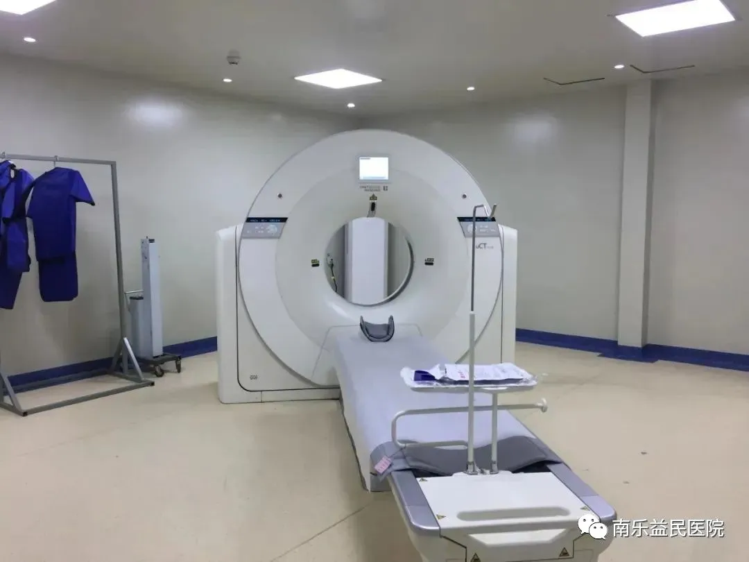
Our Hospital’s Advanced Equipment
The 64-slice 128-layer spiral CT purchased by our hospital is known as the “missile destroyer of the CT world”. It has high technological content, complete functions, and can examine various parts of the body. Compared with 16-slice and 32-slice CTs, the 64-slice 128-layer spiral CT is more suitable for examinations related to the heart, lungs, and blood vessels, and can be used to screen for coronary heart disease, lung cancer, liver cirrhosis, as well as analyze benign and malignant tumors. When examining the coronary arteries of the heart, doctors can see complete and clear three-dimensional images of the coronary arteries without cutting the skin or inserting catheters, while significantly reducing the amount of contrast agent used, making it quicker, more comfortable, and safer.
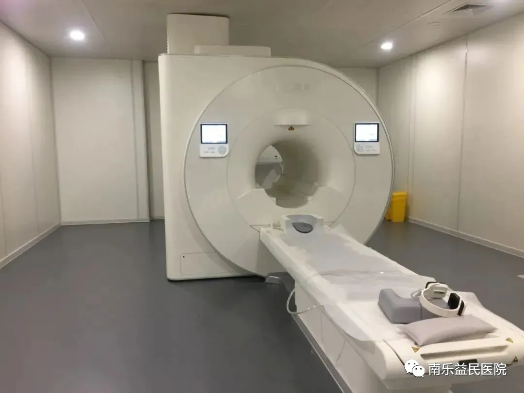 3.0 superconducting magnetic resonance, this device is one of the most advanced medical devices in the world today. It integrates research and clinical functions, providing higher image quality, richer examination methods, faster examination speeds, and more examination content. It guarantees early detection, early diagnosis, and early treatment of diseases. This device has significant advantages in magnetic resonance imaging examinations of all parts of the body! Especially in the areas of the nervous system, bones and joints, muscles, abdomen, blood vessels, breast, adnexa, prostate, and whole-body tumor screening. The clinical application prospects are broader, significantly enhancing the comprehensive strength of our hospital’s medical imaging department. It promotes the standardized development of regional medical imaging in Nanle County, providing clear images that offer accurate diagnostic bases for clinical diagnosis, addressing practical issues such as long appointment waiting times.
Emergency phone numbers:
0393-6788120
0393-6789120
Health check appointment phone:
0393-6888881