Life is accompanied by various stresses, and stress responsiveness is one of the essential characteristics of living organisms. The brain serves as the central hub for vertebrates to perceive and regulate stress, yet there remains a lack of clear understanding regarding how neurons initially sense and transduce stress signals at the cellular level. Research in this area aids in understanding the cellular mechanisms of neuronal stress responses and provides new theories for the prevention and treatment of neuropsychiatric disorders such as anxiety and depression.
Primary cilia are small cellular structures based on microtubules that protrude from the surface of non-dividing cells in vertebrates. They are considered the cell’s antenna, sensing extracellular signals and regulating cellular behavior and function through downstream pathways. Primary cilia structures were discovered in mammalian cortical neurons over 60 years ago, yet there is still a lack of understanding regarding their recognized extracellular signals and signaling pathways.
On March 7, 2025, the Shisonghai Research Group from the School of Life Sciences at Tsinghua University, the Tsinghua-IDG/McGovern Institute for Brain Research, the Beijing Frontier Research Center for Biological Structures, and the New Cornerstone Science Laboratory published a research article titled Primary ciliary protein kinase A activity in the prefrontal cortex modulates stress in mice in the journal Neuron. This study reveals a new mechanism by which primary cilia on excitatory neurons in the mouse prefrontal cortex regulate animal stress for the first time.
The study observed the morphology of primary cilia in brain neurons and found that two classic animal stress models—chronic restraint stress (CRS) and chronic social defeat stress (CSDS)—both led to a specific increase in primary cilia in the prefrontal cortex (PFC), a brain region associated with stress, suggesting a correlation between primary cilia and animal stress. To further analyze this correlation, the research team specifically knocked out the key protein CEP83 that regulates primary cilia formation in excitatory neurons of mature mouse cortex using the Cre/LoxP system, creating a primary cilia-deficient model. Cilia-deficient mice exhibited normal cortical structure, social behavior, and learning and memory functions, but showed a significant decrease in anxiety levels and acute stress response. Furthermore, chronic stress induced depressive-like behavior in wild-type mice, while cilia-deficient mice displayed resistance to chronic stress, exhibiting antidepressant-like characteristics. To further confirm the importance of primary cilia function in stress regulation in mice, the research team specifically knocked out another key protein for primary cilia assembly, TTBK2, creating a second cilia-deficient mouse strain. In these mice, a significant decrease in acute and chronic stress response levels was also observed, further indicating that primary cilia in neurons are involved in the perception and regulation of animal stress.
To elucidate the functional mechanisms of neuronal primary cilia, the research team found that the stress hormone corticosterone could similarly induce a significant increase in primary cilia in PFC neurons, while chronic corticosterone injection-induced depressive-like behavior and reduced excitability in PFC excitatory neurons were dependent on the integrity of primary cilia. To determine the core role of the PFC, the research team selectively knocked out primary cilia in excitatory neurons of the PFC using recombinant adeno-associated virus, replicating the changes in stress-related behavior in mice, while knocking out primary cilia in the sensory cortex did not alter the relevant behaviors in mice. Finally, the team discovered that corticosterone could cause an increase in cAMP levels within the primary cilia of PFC neurons using a cAMP probe localized to primary cilia, and specific inhibition of the downstream protein kinase A (PKA) signaling pathway of cAMP led to a decrease in stress response and antidepressant-like behavior in mice. These results indicate that primary cilia in excitatory neurons of the mouse PFC perceive the stress hormone corticosterone through the cAMP/PKA signaling pathway, mediating the regulation of neuronal activity related to stress and affecting behavioral phenotypes.
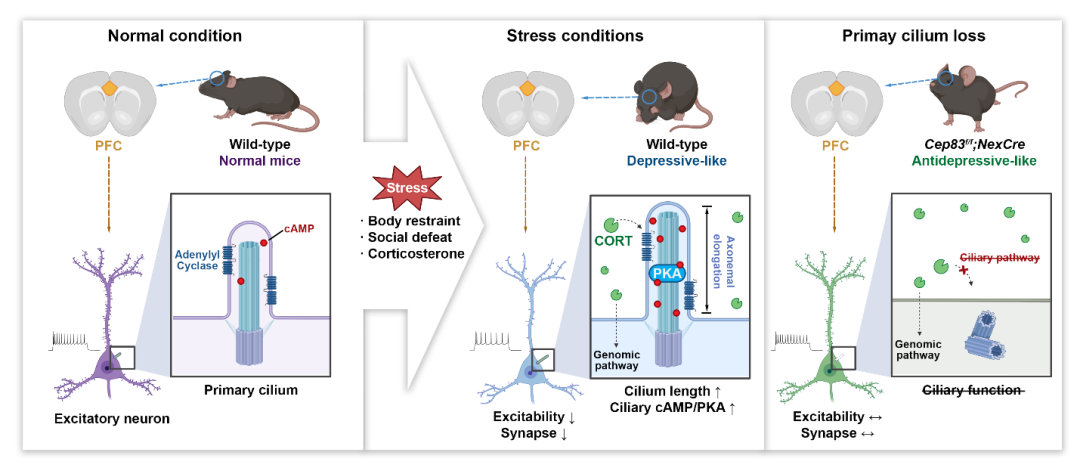
This research fills the gap in understanding the function of primary cilia in mature cortical neurons and opens new directions for understanding the processes of animal stress perception and the formation of depressive-like behaviors.
Dr. Yang Jiajun, a “Shuimu Scholar” from Tsinghua University, is the first author of this article, while Professor Shisonghai from the School of Life Sciences at Tsinghua University is the corresponding author. Researcher Shi Hang from the School of Life Sciences at Tsinghua University and the Center for Structural Biology High-Precision Innovation is a co-corresponding author. The Biomedical Optics Center team from the Shenzhen Institutes of Advanced Technology, Chinese Academy of Sciences, made significant contributions to this research.
Original link:
https://doi.org/10.1016/j.neuron.2025.02.002
Editor: Eleven
Academic Collaboration Organization
(*Rankings are not in order)
Strategic Partners
(*Rankings are not in order)
(*Rankings are not in order)
··
Reprint Notice
[Original Article] BioArt original article, personal forwarding and sharing are welcome, but reprinting without permission is prohibited. All works published are owned by BioArt. BioArt reserves all legal rights, and violators will be prosecuted.

BioArt

Med

Plants

Talent Recruitment

Conference Information
Recent Live Recommendations