01 Forum Introduction
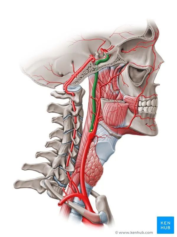
Internal carotid artery occlusion (ICAO) is one of the important factors leading to ischemic stroke. Based on the onset speed, it can be divided into acute internal carotid artery occlusion (AICAO) and chronic internal carotid artery occlusion (CICAO). Typically, occlusion lasting more than 4 weeks is classified as CICAO, while occlusions within 1 week are classified as AICAO. Patients with occlusions lasting 1 to 4 weeks are less common in clinical practice and have no clear classification. Most patients are diagnosed with AICAO due to acute ischemic stroke (AIS), while many CICAO patients show no significant clinical symptoms in the early stages.
02 Epidemiological Characteristics
Paciaroni et al. reported that AICAO accounts for 6% to 15% of AIS events; Waqas et al.’s study included 2245 AIS patients, of whom 113 were diagnosed with AICAO, with a prevalence of 5.0%. Some studies have reported that the incidence of symptomatic CICAO among Caucasians in the United States is 6 per 100,000. Verlato et al.’s research examined 1433 patients with atherosclerosis, carotid bruits, and specific or non-specific neurological symptoms using Doppler ultrasound to assess carotid and periorbital blood flow, finding 41 cases of CICAO, of which 19 (46.3%) were symptomatic and 22 (53.7%) were asymptomatic.
03 Etiology
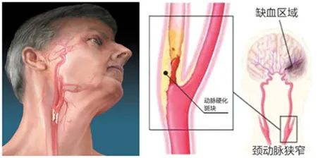
Atherosclerosis is the most common cause of ICAO, often occurring at the origin of the artery. Damage to the vascular endothelium can gradually progress to atherosclerosis and form plaques, leading to significant narrowing of the vascular lumen and eventually evolving into CICAO; once a plaque ruptures and causes bleeding, it activates and aggregates platelets, potentially leading to local vascular embolism or distal vascular embolism caused by plaque detachment, which is related to AICAO.
ICAO caused by carotid artery dissection is common at a location 2 to 3 cm from the bifurcation of the common carotid artery, resulting from genetic factors, excessive neck movement, infection, etc., causing damage to the internal carotid artery intima and potentially leading to intimal tear. Blood entering the arterial wall creates a hematoma within the layer, causing the internal carotid artery lumen to narrow or even occlude; the speed of intimal tear is critical for the acute or chronic evolution of occlusion. Other causes of AICAO include cardiogenic embolism, pituitary stroke, and carotid surgery. Among these, cardiogenic embolism is one of the main causes of AICAO, with atrial fibrillation being the most common, often embolizing to the upper segment or intracranial segment of the internal carotid artery.
Causes of CICAO also include end-stage moyamoya disease, fibromuscular dysplasia, cerebral aneurysms, trauma, Takayasu arteritis, giant cell arteritis, systemic lupus erythematosus, Sjögren’s syndrome, Kimmell’s disease, and moyamoya syndrome that has reached the stage of internal carotid artery occlusion.
04 Pathophysiology
The pathophysiological changes of AICAO mainly include two points: firstly, the platelet emboli and in situ thrombus at the AICAO site lead to isolated internal carotid artery occlusion or downstream serial occlusion due to combined atherosclerotic plaques, resulting in severe symptomatic stroke; secondly, hemodynamic changes secondary to AICAO lead to a rapid decrease in distal arterial perfusion pressure, and due to the rapid onset, collateral compensation is poor, severe hemodynamic disturbances can result in large area infarction and edema in the occluded hemisphere.
The pathophysiological changes of CICAO mainly include three points:
-
Firstly, residual thrombus or plaque detachment at the occlusion site circulates downstream, as there is better blood flow compensation, it can manifest as recurrent transient ischemic attacks (TIA);
-
Secondly, hemodynamic changes secondary to CICAO are milder than those of AICAO, with watershed infarcts between the anterior cerebral artery (ACA) and middle cerebral artery (MCA) and between MCA and posterior cerebral artery (PCA) being more common;
-
Thirdly, the chronic development of ICAO leads to dilation of distal collateral vessels and continuous formation of new anastomoses, such as connections between the meningeal collateral vessels and cortical capillaries, and anastomoses between the external carotid artery and branches of the internal carotid artery, as well as secondary branches formed between meningeal collateral vessels and ACA-MCA, MCA-PCA.
Connolly et al. believe that after ICAO occurs, the anterior communicating artery and posterior communicating artery are the main collateral compensatory vessels, while the retrograde ophthalmic artery, meningeal collateral vessels, and PCA are secondary collateral compensatory vessels. The study found that 63% (71/113) of CICAO patients had retrograde ophthalmic arteries, which often indicates severe hemodynamic disturbances.
05 Clinical Manifestations
AICAO often manifests as AIS due to the inability to quickly form good collateral compensation, and its specific clinical manifestations are related to AICAO and the sites of associated occlusion. Proximal embolism of the ophthalmic artery is mainly seen in CICAO patients, while downstream serial occlusion mainly involves ACA and MCA, and can also embolize distal branches of PCA through the embryonic-type PCA.
A typical occlusion of the main trunk of one MCA can cause contralateral limb weakness, contralateral visual field defects, contralateral sensory numbness, and eye gaze. If the dominant hemisphere’s brain function is severely impaired, complete aphasia may occur, while progression of non-dominant hemisphere infarction can lead to unilateral neglect symptoms. Occlusion of the penetrating arteries of MCA usually leads to pure motor, sensory-motor, dysarthria, and ataxia. The specific clinical manifestations of ACA occlusion include limb weakness, with the lower limbs more affected than the upper limbs, as well as other symptoms such as motor aphasia, apraxia, urinary incontinence, amnesia, drowsiness, and Parkinsonian symptoms.
CICAO has a longer course, with dizziness and recurrent TIAs being common. If there is sufficient collateral circulation compensation, there may be no significant clinical manifestations. Embolism of the ophthalmic artery or poor blood flow compensation may manifest as recurrent transient retinal ischemia, and some patients may develop chronic ocular ischemia syndrome. If the perfusion pressure in watershed areas secondary to CICAO drops significantly, clinical symptoms similar to AICAO may also appear. Other rare clinical manifestations of CICAO include involuntary unilateral limb tremors, syncope, headache, facial pulsation, and cognitive decline.
06 Imaging Examination and Assessment
Ultrasound examination of the head and neck mainly includes transcranial Doppler ultrasound and carotid ultrasound. Transcranial Doppler ultrasound can reflect the retrograde flow of the ophthalmic artery and is of great significance for assessing intracranial hemodynamics. Although the accuracy of ultrasound in assessing vascular occlusion is lower than that of CT angiography (CTA), MR angiography (MRA), and DSA, it has advantages in showing the characteristics of vessels and thrombi at the occlusion site.
Ventura et al. proposed using second-generation contrast-enhanced ultrasound (CEUS) as the gold standard for diagnosing ICAO. They compared the diagnosis of ICAO using conventional Doppler ultrasound, CTA, and CEUS, finding that the false positive rate of conventional Doppler ultrasound was as high as 26.9% (21/78), while the false positive rate of CEUS was 2.6%, with a sensitivity of 100.0%, specificity of 90.5%, negative predictive value of 100.0%, positive predictive value of 96.6%, and accuracy of 97.4%. Moreover, the microbubble contrast agent used in CEUS is relatively safe and suitable for patients with renal dysfunction who cannot use iodine contrast agents.
MRI is widely used in neurological examinations, with common sequences for arterial imaging including time of flight (TOF) and contrast-enhanced MRA. Recent studies involving 131 consecutively admitted AIS patients undergoing MRI showed that TOF-MRA had a higher diagnostic rate for arterial occlusion [48.1% (63/131) compared to 39.7% (52/131)], but also had a higher false positive rate [20.6% (13/63) compared to 1.9% (1/52)], and was less precise in measuring thrombus length than contrast-enhanced MRA [22% (14 cases) compared to 75% (38 cases)]; additionally, for intracranial vascular occlusion, the diagnostic accuracy of TOF-MRA was 89%, while that of contrast-enhanced MRA reached 99%, and contrast-enhanced MRA was more accurate in determining the location and length of thrombi. High-resolution MRI is gradually being widely used for ICAO examinations, providing high value in assessing atherosclerotic plaques and reflecting inflammatory responses within the plaques.
CTA is specific for assessing atherosclerotic plaques. Michel et al. reported that the “carotid ring sign” on CTA images can distinguish AICAO from CICAO, but further research is needed for validation. A new type of four-dimensional CTA technology improves the accuracy of diagnosing vascular lesions by analyzing collected CTA and CT perfusion data while reducing artifacts. DSA remains the gold standard for diagnosing cerebrovascular diseases, allowing observation of blood flow direction, collateral compensation, and completing an integrated approach from examination to treatment. However, due to its invasiveness and high cost, it has not become the first-choice diagnostic method for most patients.
07 Treatment Methods for AICAO
Treatment for AICAO patients mainly includes endovascular thrombolysis, endovascular therapy, and carotid endarterectomy (CEA). Among these, endovascular thrombolysis includes intravenous thrombolysis and intra-arterial thrombolysis, but in clinical practice, intravenous thrombolysis is the primary method. Endovascular therapy includes balloon dilation, stent placement, thrombectomy, and catheter aspiration.
2.1.1 Intravenous or Intra-arterial Thrombolysis: For AICAO patients presenting with AIS symptoms within 4.5 hours, intravenous thrombolysis remains the preferred treatment. Saqqur et al. found that AICAO patients with undetectable residual blood flow signals and those with occlusion at the terminal internal carotid artery had a low rate of recanalization after intravenous thrombolysis. A retrospective study included 100 isolated AICAO patients, and the recanalization rate after intravenous thrombolysis was 54%, with a mortality rate of 21%, and 12% of patients experienced symptomatic intracranial hemorrhage, while 27% had a modified Rankin Scale (mRS) score of 0-1 at three months.
The internal carotid artery occlusion (ICARO) trial included 506 AICAO patients divided into an intravenous thrombolysis group and a conservative treatment group, with 253 patients in each group. The results showed that the proportion of patients with good prognosis (mRS score ≤ 2) was higher in the intravenous thrombolysis group [28.9% (73/253) compared to 20.6% (52/253), OR=1.80, P=0.037], but the mortality rate was higher [25.7% (65/253) compared to 15.4% (39/253), OR=2.28, P=0.001], and the incidence of fatal intracranial hemorrhage was higher, although the difference was not statistically significant [2.8% (7/253) compared to 0.4% (1/253), OR=7.17, P=0.068].
Simple intra-arterial thrombolysis and endovascular therapy are both invasive procedures, but endovascular therapy has higher recanalization rates and more favorable outcomes. Kappelhof et al. summarized and analyzed 32 studies on AICAO treatment, including 1107 patients. In patients with occlusion of the intracranial segment of the internal carotid artery, endovascular therapy had a higher recanalization rate compared to intra-arterial thrombolysis (69% vs. 38%, P<0.01) and more favorable outcomes (mRS score 0-2, 34% vs. 12%, P<0.01); in patients with occlusion of the extracranial segment of the internal carotid artery, endovascular therapy also had a higher recanalization rate (87% vs. 48%, P=0.01), more favorable outcomes (68% vs. 38%, P<0.01), and a lower mortality rate (18% vs. 41%, P=0.048).
2.1.2 Endovascular Therapy or Combined Endovascular Therapy with Intravenous Thrombolysis: Compared to intravenous thrombolysis, endovascular therapy is more beneficial for improving functional prognosis. Mokin et al. systematically reviewed the treatment effects of intravenous thrombolysis and endovascular therapy, defining favorable outcomes as mRS score ≤ 2 three months later. The results showed that patients receiving endovascular therapy (584 patients) had more favorable outcomes than those receiving only intravenous thrombolysis (385 patients) [33.6% (196/584) vs. 24.9% (96/385), P=0.004], although there was a higher incidence of symptomatic intracranial hemorrhage [11.1% (65/584) vs. 4.9% (19/385), P=0.001], it did not lead to an increase in overall mortality [32.0% (187/584) vs. 27.3% (105/385), P=0.12].
However, the ICARO-3 trial compared the efficacy differences between the endovascular therapy group and the intravenous thrombolysis group, with 324 patients in each group. The proportion of patients with an mRS score of 0-2 at three months was higher in the endovascular therapy group [32.4% (105/324) vs. 27.4% (89/324); OR=1.25, P=0.10], and the mortality rate was lower [17.6% (57/324) vs. 23.1% (75/324); OR=0.71, P=0.07], but the differences were not statistically significant; however, the incidence of intracranial hemorrhage was higher [37.0% (120/324) vs. 17.3% (56/324); OR=2.82, P=0.001], and the incidence of fatal intracranial hemorrhage was also higher [5.9% (19/324) vs. 2.2% (7/324); OR=3.31, P=0.01], thus this study did not consider endovascular therapy to be superior.
Intravenous thrombolysis combined with endovascular therapy provides a further treatment option for patients for whom intravenous thrombolysis is ineffective. Anadani et al. noted that intravenous thrombolysis combined with endovascular therapy did not increase the risk of bleeding complications compared to endovascular therapy alone. In a study involving 205 patients with tandem AICAO, 125 underwent intravenous thrombolysis combined with endovascular therapy, while the remaining 80 underwent endovascular therapy alone. Compared to the endovascular therapy group, the combined therapy group had a lower mortality rate at 90 days [8% (10/125) vs. 20% (16/80), P=0.017], and the difference in the incidence of postoperative symptomatic intracranial hemorrhage was not statistically significant [5% (6/125) vs. 8% (6/80), P=0.544], while the difference in favorable outcomes (mRS score ≤ 2) at three months postoperatively was not statistically significant [62% (76/123) vs. 51% (40/79), P=0.145].
Systematic reviews by Romoli et al. also support this bridging treatment, showing that the bridging treatment group had better favorable outcomes compared to the intravenous thrombolysis group [mRS score ≤ 2, 46.4% (26/56) vs. 28.0% (115/410), OR=2.2, 95%CI: 1.3-3.7], a higher incidence of symptomatic intracranial hemorrhage [25.0% (14/56) vs. 6.1% (25/410), OR=5.1, 95%CI: 2.5-10.5], but no increase in mortality [19.6% (11/56) vs. 25.1% (103/410), OR=0.7, 95%CI: 0.4-1.4]; the bridging treatment group also had more favorable outcomes compared to the endovascular therapy group alone [mRS score ≤ 2, 46.4% (26/56) vs. 31.0% (45/145), OR=1.9, 95%CI: 1.1-3.4], and a higher recanalization rate [94.4% (17/18) vs. 46.2% (12/26), OR=19.8, 95%CI: 7.7-51.4].
Zi et al.’s latest prospective study originally planned to include 970 patients with acute large vessel occlusion, but the study was prematurely terminated due to efficacy concerns. Among the 234 patients already included, endovascular therapy did not show statistically significant differences in functional independence [mRS score ≤ 2, 54.3% (63/116) vs. 46.6% (55/118)], symptomatic intracranial hemorrhage [6.1% (7/115) vs. 6.8% (8/117)], and 90-day mortality [17.2% (20/116) vs. 17.8% (21/118)] between the endovascular therapy and intravenous thrombolysis combined with endovascular therapy groups (all P>0.05), suggesting the non-inferiority of direct endovascular therapy and indicating that endovascular therapy could be performed directly without going through the thrombolysis process. However, this study had a small proportion of AICAO subtypes, with 18 cases (15.5%) and 17 cases (14.4%) in the two groups, respectively, and further large-scale studies are needed to validate this conclusion in AICAO patients.
2.1.3 Endovascular Therapy Combined with CEA: Hasegawa et al. reported three AICAO patients who underwent combined endovascular therapy and CEA, with NIHSS scores of 26, 22, and 19 upon admission, and successful recanalization of occluded vessels after combined surgery, with NIHSS scores of 3, 9, and 2 one month postoperatively.
Schubert et al. reported that all 12 AICAO patients who underwent combined surgery had successful recanalization of occluded vessels, and 7 patients had good prognoses (mRS score ≤ 2) at three months postoperatively. Although combined endovascular therapy and CEA can treat AICAO from an etiological perspective, the longer surgical time increases the risk of acute neurological dysfunction, making this combined surgery more applicable to CICAO patients.
08 Treatment Methods for CICAO
2.2.1 Pharmacological Treatment: CICAO patients without obvious clinical manifestations do not require special treatment, but it is important to control risk factors associated with atherosclerosis; for symptomatic CICAO patients, immediate secondary stroke prevention should be initiated, and in the absence of clinical contraindications, low-dose aspirin (75-150 mg/d) combined with clopidogrel (75 mg/d) should be actively used for antiplatelet therapy. Dual antiplatelet therapy should be switched to single antiplatelet therapy after three months and maintained long-term, during which lipid-lowering therapy can further reduce the occurrence of adverse cardiovascular and cerebrovascular events.
Studies have shown that the maximum benefit of dual antiplatelet therapy occurs within 21 days after TIA or AIS, and it is recommended to switch to single antiplatelet therapy after three weeks. A new meta-analysis showed that long-term clopidogrel monotherapy is more effective than aspirin monotherapy. The analysis included 29,357 recent ischemic stroke patients, with 14,293 receiving clopidogrel alone and 15,064 receiving aspirin alone for secondary stroke prevention, showing that the risk of major cardiovascular and cerebrovascular adverse events was significantly lower in the clopidogrel group compared to the aspirin group (RR=0.72, 95%CI: 0.53-0.97).
2.2.2 Vascular Reconstruction: For CICAO patients with significant hemodynamic disturbances who are ineffective with medical treatment, vascular reconstruction and recanalization procedures may be performed. Following the treatment methods for moyamoya disease, vascular reconstruction can be classified into direct vascular reconstruction, indirect vascular reconstruction, and mixed vascular reconstruction. Standalone indirect vascular reconstruction is represented by the encephalo-myo-synangiosis technique, which currently lacks consensus on its classification as a primary treatment for CICAO, but may be used for patients who cannot undergo direct vascular reconstruction and recanalization treatment. Intracranial-extracranial bypass grafting is a classic direct vascular reconstruction technique, but its efficacy and safety remain controversial.
The carotid occlusion surgery study (COSS) included 195 patients with atherosclerotic CICAO, of which 97 underwent bypass grafting and 98 received medical treatment. The stroke incidence on the same side within 30 days was higher in the surgical group than in the medical group [14.4% (14/97) vs. 2.0% (2/98)], and at the two-year follow-up, the difference in the occurrence of primary endpoint events between the surgical and medical groups was not statistically significant (21.0% vs. 22.7%, P=0.78). Recent results from COSS again denied the benefit of bypass grafting in recurrent hemispheric stroke patients, with the trial also including 195 patients with CICAO caused by atherosclerosis, with 100 cases in the recurrent hemispheric ischemia (rHEMI) group and 50 in each of the surgical and medical groups; 95 cases in the non-rHEMI group with 47 in the surgical group and 48 in the medical group. Defining ipsilateral ischemic stroke as the primary endpoint and following up for two years, the results showed that there were no statistically significant differences in the primary endpoint between the surgical and medical treatments, both in the rHEMI group (26.3% vs. 22.4%, P=0.66) and in the non-rHEMI group (18.9% vs. 19.5%, P=0.94).
The superficial temporal artery-middle cerebral artery branch anastomosis combined with encephalo-myo-synangiosis is currently the recommended mixed vascular reconstruction technique, widely applied in moyamoya disease patients. Han Hongyan et al. retrospectively analyzed 109 moyamoya disease patients who underwent mixed vascular reconstruction (184 vessels), finding that during the follow-up period, 5 patients experienced recurrent TIA, and 5 patients had cerebral hemorrhage; all 10 patients had Suzuki stages between 5 and 6. This study indicated that mixed vascular reconstruction is not effective for end-stage moyamoya disease patients. Additionally, several reports suggest that end-stage moyamoya disease patients with severe intracranial vascular lesions cannot benefit from mixed vascular reconstruction.
The authors believe that atherosclerotic CICAO and end-stage moyamoya disease patients often have severe intracranial vascular lesions, leading to poorer prognosis, but this does not negate the efficacy of vascular reconstruction in CICAO patients of other etiologies. Furthermore, when assessing endpoint events for surgical efficacy, more aspects can be considered, and specific conclusions require analysis of etiology and prognostic factors in a large sample of CICAO patients undergoing vascular reconstruction.
2.2.3 Recanalization Procedures: Recanalization procedures include CEA, endovascular therapy, and combined CEA with endovascular therapy. CEA is suitable for short occlusions at the origin of the internal carotid artery. Liang Xiaolong et al. reported 23 CICAO patients with proximal occlusion, showing 100% successful recanalization after CEA, and at three-month follow-up, only one asymptomatic patient prior to surgery experienced reocclusion of the internal carotid artery on the surgical side, while the others did not experience complications such as hyperperfusion syndrome, intracranial hemorrhage, or incision infection.
Moreover, endovascular therapy can achieve high success rates for occlusions below the upper segment of the internal carotid artery. Chen et al. conducted a retrospective analysis of 138 CICAO patients, showing an overall recanalization rate of 61.6% (85/138), with recanalization rates of 93% (26/28), 80% (20/25), 73% (22/30), 33% (9/27), and 29% (8/28) for occlusions at the petrous segment and below, cavernous segment, tuberculum sellae segment, ophthalmic segment, and communicating segment, respectively.
For long segment occlusions from the origin of the internal carotid artery to above the petrous segment, combined CEA and endovascular therapy have high recanalization rates. Zhang et al. reported that in 65 CICAO patients with long segment occlusions, 35 received medical conservative treatment, while 30 underwent combined surgical treatment, achieving a 100% patency rate in the occluded segment distal to the surgical site, with neurological function improvement in 26 patients. Among the perioperative complications, one patient experienced recurrent laryngeal nerve injury, and one had a microguidewire perforation causing intracranial hemorrhage. Three months after surgery, angiographic results showed patency in all occluded vessels; the surgical group showed significant improvement in mRS scores at three months [(2.5±0.6) vs. (3.4±0.6), P<0.01], while the conservative treatment group showed no significant improvement [(3.4±0.7) vs. (3.5±0.8), P>0.05].
Currently, there are no guidelines clearly indicating the preference between vascular reconstruction and recanalization procedures for CICAO patients. Clinical physicians should comprehensively consider specific etiology, surgical difficulty, advantages and disadvantages of the surgery, and the operator’s proficiency when deciding on a surgical approach.
09 Summary
When clinically diagnosing and treating ICAO, it is essential to clarify the duration of occlusion and the speed of onset, and to choose an individualized treatment strategy. However, due to the many correlations between AICAO and CICAO, especially when CICAO patients experience AIS, treatment strategies should also incorporate experiences from AICAO treatment. Domestic research on ICAO still lacks large-sample long-term follow-up studies, particularly for CICAO patients, where long-term follow-up is crucial. Improving patient follow-up data can provide data references for the research of new treatment protocols.
Source: Yang Cheng, Long Jiang. Advances in Research on Internal Carotid Artery Occlusion [J]. Chinese Journal of Cerebrovascular Diseases, 2021, 18(03): 210-216.
More Exciting Reads
Essential Tremor Is More Than Just Shaking!
Paradoxical Embolism
A Kaleidoscope of Neurological Diseases!
The Future of Behavioral Sleep Medicine
How to Diagnose and Treat Restless Legs Syndrome (RLS)? | Latest Guidelines