

Journal: Nature Communications
Impact Factor: 14.7
Main Technology:scRNA-seq
Introduction
Lymphocyte receptors have evolved independently in jawed and jawless vertebrates, exhibiting similar adaptive immune responses. However, the functional subtypes and molecular diversity of lymphocytes in jawless vertebrates remain poorly defined compared to jawed animals. The authors analyzed the gills, intestines, and blood of the lamprey (Lampetra morii) using scRNA-seq and employed a full-length transcriptome as a reference. The results indicated that T-like cells exhibited greater tissue-specific heterogeneity compared to B-like cells. The authors identified a unique T-like cell subtype expressing the homolog MPL-L, a receptor for non-lymphoid hematopoietic growth factors. These MPL-L+ T-like cells displayed characteristics distinct from T cells in jawed vertebrates, particularly with elevated expression of hematopoietic genes. Furthermore, MPL-L+VLRA+ T-like cells were found to be widely present in the lamprey’s skin, gills, liver, kidneys, and intestines, proliferating under the influence of T cell mitogens and recombinant human thrombopoietin. These findings provide new insights into the adaptive immune responses of jawless vertebrates and offer new perspectives on the evolution of adaptive immunity.
Main Technology
scRNA-seq
Main Results
1. Multi-immune cell atlas of L. morii larvae
The adaptive immune system (AIS) of jawless and jawed vertebrates is supported by a unique lymphocyte receptor system. The authors utilized the full-length transcriptome of L. morii for further analysis of single-cell RNA sequencing data. The results demonstrated that the data provided by the full-length transcriptome was sufficient to support subsequent analyses.
To investigate the diversity of cell types in the immune tissues of lampreys, single-cell samples were collected from three tissue types: gills, blood, and intestines (including mesentery), resulting in six gill libraries, three blood libraries, and five intestinal libraries. A total of 25,978 gill cells, 16,483 blood cells, and 23,204 intestinal cells were retained for further analysis, with median counts of 590, 489, and 873, respectively. Erythroid and immune cell populations were distinguished based on ALAS1 and CD45. T-like and B-like cells were further differentiated based on the expression of VLRA, VLRC, and VLRB genes. The cell atlas of the gills was first annotated, focusing on identifying myeloid cell populations and analyzing differentially expressed genes through previous literature. Notably, a specific cell population was identified characterized by the expression of various interferon-stimulated genes (ISGs), including MX1, TRIM25/29/80, TRIM65, etc. This expression pattern suggests that these ISG-expressing cells have a unique response to interferon signaling. A hematopoietic cell population (TAL1, ITGB3, PMP22, etc.) was also identified, and the aforementioned marker genes were used for subsequent annotations of the intestinal and blood atlases.
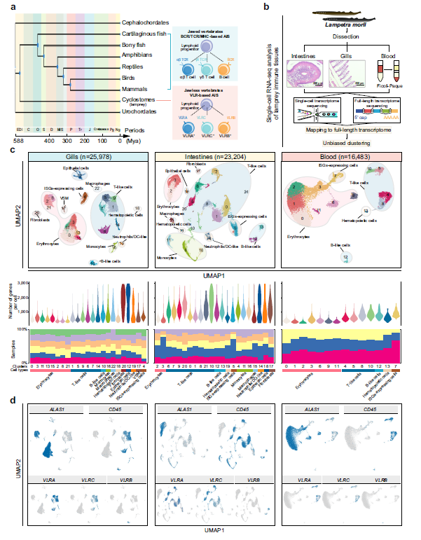
Fig 1. Transcriptional characteristics of gill, intestinal, and blood atlases
2. Inter-tissue cell heterogeneity
To better compare the differences in cell types among gills, blood, and intestines, these cell types were integrated and clustered, including erythrocytes, ISG-expressing cells, hematopoietic cells, endothelial cells, fibroblasts, monocytes, macrophages, neutrophils/DC cells, epithelial cells, T cells, and B cells. The proportion of erythrocytes and lymphocytes was higher than that of other cell types in the other three tissues. The concentration of innate immune cells in the intestines was relatively higher than in the gills and blood. VLRA+ cells in gill filaments and gill slices were identified using immunohistochemical methods with anti-VLRA pAb. Results showed a significant amount of brown positive signals observed in all studied tissues.
Further MetaNeighbor analysis was conducted to explore the relationships of cell types among the three tissues. ISG-expressing cells, hematopoietic cells, and erythrocytes exhibited similar characteristics in the gills and intestines, showing the same expression patterns across these three different tissues. Significant cross-tissue similarity was observed in B cells, but not in T cells. The gene expression profiles of B cells showed significant consistency across all three tissues. However, certain T cell markers and differentially expressed genes exhibited significant heterogeneity, attributable to the tissue-specific differentiation of T cell subtypes.
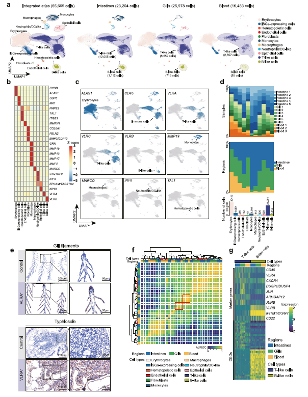
Fig 2. Integrated analysis of gill, intestinal, and blood atlases
3. Discovery and characterization of different lymphocyte subtypes
T cells and B cells were re-integrated and analyzed. Based on differential gene expression profiles, five T cell subtypes and one B cell population were identified. Analysis of characteristic genes revealed that most T cell subtypes highly expressed genes similar to traditional T cell functions. The hallmark genes of MPL-L+T cells are related to hematopoiesis. The MPL-L gene is highly and widely expressed in MPL-L+T cells and co-expressed with VLRA. Interestingly, MPL is a receptor for non-lymphoid hematopoietic growth factors in jawed vertebrates, primarily binding to its ligand thrombopoietin (TPO), involved in the self-renewal of hematopoietic stem cells (HSCs) and the maturation of megakaryocyte-derived platelets, but not in the development and activation of lymphocytes. Meanwhile, MPL can activate a range of signaling pathways, including JAK/STAT phosphorylation pathways and RAS pathways. However, current mouse studies indicate that introducing the MPL gene into T cells via transgenic technology can enhance T cell self-renewal and anti-tumor capabilities, based on the overlap of the TPO-MPL signaling pathway with co-stimulatory molecules and cytokine production during T cell activation. Based on the observed transcriptional heterogeneity and unique distribution patterns, the authors propose that MPL-L+T cells constitute an unconventional population within the lamprey lymphocyte lineage.
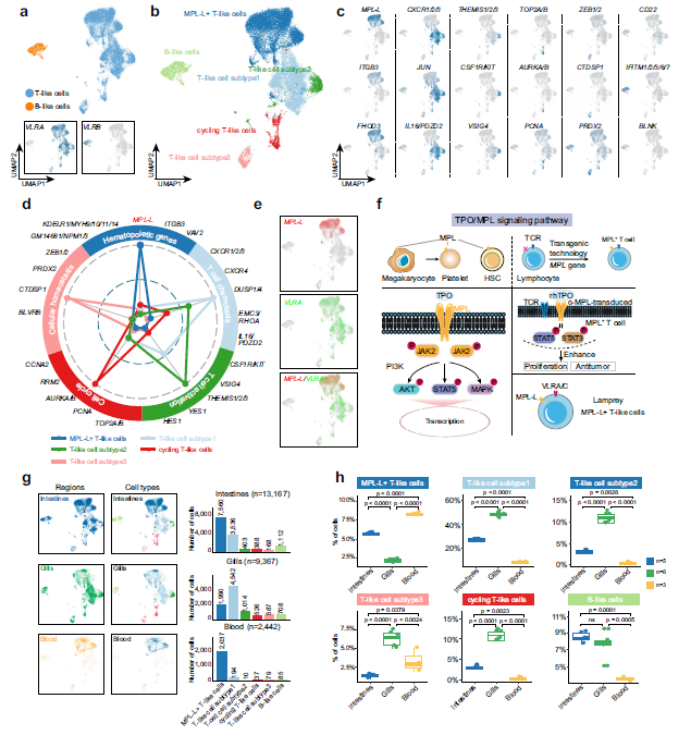
Fig 3. Transcriptional characteristics and distribution of lymphocyte subpopulations
4. MPL-L+T cells represent a unique aspect of the adaptive immune system in jawless vertebrates, differing from jawed vertebrates
SAMap analysis results showed extensive consensus between lampreys and zebrafish and mice regarding innate immune cells and hematopoietic cells/hematopoietic stem cells/megakaryocytes. B cells showed lower similarity to B cells in mice, while significant B cell consistency was observed in zebrafish. In the cross-species mapping of the lamprey T cell lineage, complex relationships were revealed. The authors analyzed the distribution and expression of mpl/Mpl/MPL-L genes in zebrafish/mice and lampreys. The results indicated that the vast majority of MPL-L+T-like cells and hematopoietic cells in lampreys express the MPL-L gene, while in zebrafish/mice, only HSCs/megakaryocytes express this gene.
To verify whether MPL-L+T-like cells are exclusive to lampreys, the authors performed in situ hybridization and co-localization analysis on multiple organs of zebrafish (liver, head kidney, thymus, and gills). Results showed that mpl did not co-localize with the T cell marker lck. The authors also prepared anti-Lm-MPL-L polyclonal antibodies and detected the co-localization of MPL-L with VLRA in lampreys through immunofluorescence. Co-localization of MPL-L and VLRA was observed in gill filaments, skin, kidneys, and liver. Based on these results, potential activation signals for lamprey T cells and the evolutionary differentiation of lymphocyte lineages in vertebrates were indicated.

Fig 4. Cross-species analysis of lampreys with zebrafish/mice
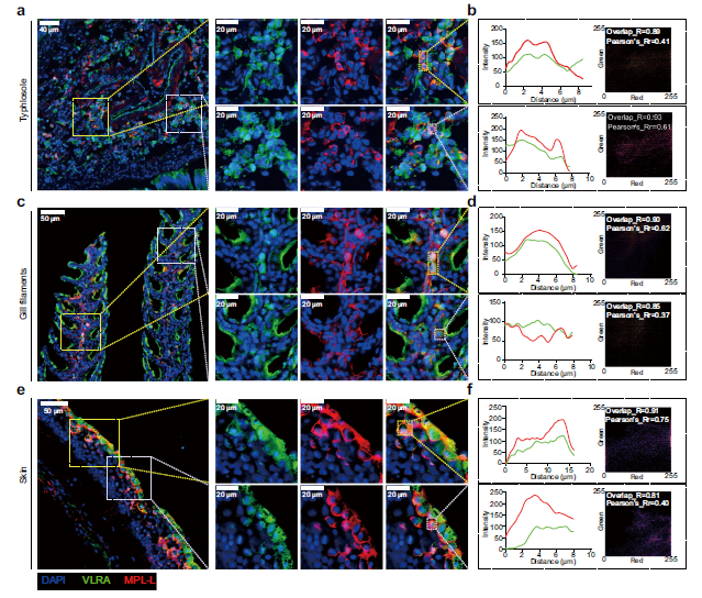
Fig 5. Double-staining immunofluorescence detection of mpl +VLRA+ cells
5. MPL-L+ VLRA+ cells respond to plant lectin-L (Pha-L) and recombinant human TPO (rhTPO)
To further verify whether MPL-L+ VLRA+ cells possess T cell antigen response characteristics and respond to human TPO, the authors injected PHA-L and rhTPO into the tail of the larvae. Immunofluorescence detection results showed that stimulation with PHA-L and rhTPO led to an increase in co-localization regions of VLRA and MPL-L, and promoted the expression of MPL-L in the mesentery. Results also indicated an increase in the proportion of MPL-L+ VLRA+ cells in gill filaments post-injection. However, no significant changes in MPL-L expression levels were observed within the MPL-L+ VLRA+ cell population in gill filaments. Additionally, flow cytometry analysis of cells stained with anti-VLRA and anti-MPL-L showed an increase in the proportion of MPL-L+ VLRA+ cells after rhTPO injection. Furthermore, PHA-L immunostimulation promoted the proliferation of VLRA+ cells and MPL+ cells. This stimulation may promote the proliferation of cells at early or late stages of differentiation, which express either MPL-L or VLRA. EdU injection also showed increased cell proliferation in gill filaments and gills after rhTPO injection.

Fig 6. rhTPO and PHA-L stimulation can lead to the proliferation of MPL-L + VLRA+ cells
6. MPL-L+T cell characteristic molecular profile and developmental trajectory
Using Monocle2 to infer state trajectories, dynamic cell transitions within T cell subpopulations were explored. Based on trajectory information, this trajectory was determined to start from T cell subtype 3, with most MPL-L+T-like cells ultimately reaching a terminal state distinct from the other three cell subpopulations. The proportion of cells expressing the VLRA gene and the average expression level of the VLRA gene were calculated for each T-like cell subpopulation. A moderate proportion of T cell subtype 3 expressed the VLRA gene, and the average expression level of the VLRA gene in T cell subtype 3 was the lowest among all T-like cell subpopulations. The expression level of the VLRA gene in T cell subtype 2 was slightly higher than in T cell subtype 3, while T cell subtypes 1 and MPL-L+T-like cells were primarily in the terminal state.
The MPL-L+T-like cell populations in the intestines, blood, and gills were distinguished through differentially expressed genes. To further confirm the heterogeneity of MPL-L+T cells in the three tissues, Monocle 2 trajectory analysis was utilized to determine the transitional states of MPL-L+T cells within these tissues. Results indicated that these cells were in unique differentiation states. Combining trajectory transition information and the proportion of cells expressing the VLRA gene, it was inferred that MPL-L+T cells exhibit transcriptional and developmental diversity within their respective tissues. During the transformation of MPL-L+T cells in the gills, blood, and intestines, three tissue-specific gene expression patterns were formed. The T cell co-stimulatory molecule CD63 and the hallmark transcription factor of innate lymphocyte lineages ZBTB16 were also expressed in MPL-L+T cells in the intestines.
To further investigate the potential role and regulatory mechanisms of the MPL-L gene in MPL-L+T cells across different tissues, the proportion of cells expressing the MPL-L gene and the average expression level of this gene were determined in three MPL-L+T cell subpopulations. In all three tissues, the MPL-L gene exhibited a significant upward trend in expression along the developmental trajectory of MPL-L+T cells. The expression of the MPL-L gene was confirmed to be positively correlated with its downstream genes JAK2 and STAT5B in MPL-L+T cells. The positive correlation in MPL-L+T cells in the intestines was more pronounced than in those in the gills and blood. These findings suggest the presence of an MPL-L-based signaling system within MPL-L+T cells, potentially coordinating the development or activation of intestinal MPL-L+T cells.
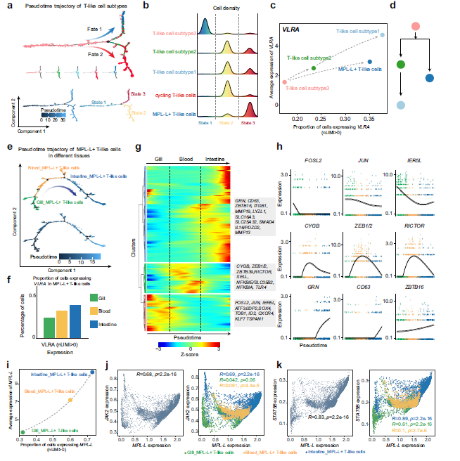
Fig 7. Transitional state analysis of MPL-L+T-like cells
References:
Huang Y, Liu X, Li S, Li C, Wang HY, Liu Q, Chen JY, Zhang Y, Li Y, Zhang X, Wang Q, Liu K, Liu YY, Pang Y, Liu S, Fan G, Shao C. Discovery of an unconventional lamprey lymphocyte lineage highlights divergent features in vertebrate adaptive immune system evolution. Nat Commun. 2024 Sep 3;15(1):7626. doi: 10.1038/s41467-024-51763-2. PMID: 39227584; PMCID: PMC11372201.
Recommended Reading
Technical Breakthrough | Secrets to Producing High-Quality Single-Cell Data from FFPE Samples
April High-Impact Literature Dispatch | Spatial Transcriptomics Series
Project Article | Cancer Cell Publishes High-Precision Single-Cell Atlas of Pan-Cancer Brain Metastasis
Conference Review | CCTB-2025 Tumor Biomarker Academic Conference
Xian Station | 2025 Berhao Bioinformatics Training Class Registration is Open!
END
This article is an original work by Berhao Bio
Feel free to share it with your friends
Please indicate that it is from Berhao Bio when reprinting
Consultation Hotline: 17702139967
Email: [email protected]


Did you discover the “share” and “like” buttons? Click here to check it out!