Accurately delivering therapeutic cells to the desired site in vivo is an emerging and promising cell therapy. However, targeting therapeutic cells to the affected area in the body can be challenging, especially when using a large number of cells, as excessive cytokine production can lead to an overactive immune response. Utilizing microrobots allows for controllable therapeutic doses of cells; however, the degradation of microrobots and real-time tracking and precise localization in vivo present significant challenges.
Recently, Dong Sun and others from City University of Hong Kong published an article titled “Development of Magnet‐Driven and Image‐Guided Degradable Microrobots for the Precise Delivery of Engineered Stem Cells for Cancer Therapy” in Small, reporting on a degradable and image-guided microrobot that achieves automatic navigation in vascular tissues for cell delivery under the drive of an external gradient magnetic field. The microrobots feature a spiky porous spherical structure and are made from synthetic composites with degradable properties, mechanical strength, and magnetic driving capabilities. By employing unique PA imaging technology, the microrobots can be visualized within tissues up to 2 cm deep. Preclinical tests conducted on nude mice with in situ liver tumors indicated that the cells released by the microrobots significantly reduced tumor growth.
1. Microrobot Design
Previous work by researchers has shown that microrobots with a spiky porous spherical structure have multiple advantages. First, besides the ease of achieving microrobot-host tissue integration and cell transfer from the robot to the tissue, the spherical structure enhances magnetic driving capability. Second, the porous spherical structure increases cell carrying capacity. Third, incorporating spiky structures into the porous spherical microrobots further enhances cell carrying capacity. The microrobots in this study adopt the same structural design but are made from degradable materials, potentially reducing immune activation and thrombosis. Polyethylene glycol (PEG) manufactured using 3D lithography technology displayed high mechanical strength and high resolution.
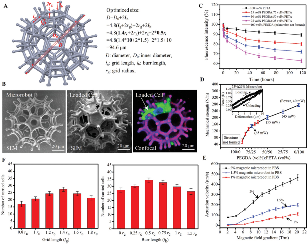
Figure 1: Design and size determination of the degradable microrobots. A) Calculating models of spiky porous microrobots with optimal sizes. B) SEM images of the microrobots (left), microrobots loaded with cells (middle), and confocal scans of GFP-MSC cultured on the microrobots (right). C) Fluorescence intensity changes over time for microrobots with different PEGDA and PETA compositions in phosphate-buffered saline (PBS). D) Mechanical strength of microrobots with different PEGDA and PETA compositions. Illustrations show mechanical tests of microrobots with 75% (volume) PEGDA: deformation of microrobots using measurable force, converting 25% (volume) PEGDA to PETA. E) Magnetic driving tests of microrobots with different compositions of 75 vol% PEGDA and 25 vol% PETA superparamagnetic particles. F) Statistical results of MSC-loaded microrobots with different mesh lengths and spike lengths.
To demonstrate that the fluorescence tracking method is suitable for visualizing the degradation process, the researchers characterized the fluorescence signals of degradable 75 vol% PEGDA: 25 vol% PETA microrobots and non-degradable 100 vol% PETA microrobots in an alkaline environment. Results indicated that the PEGDA polymer network encapsulated with fluorescent molecules can serve as an indicator of microrobot degradation. Furthermore, the researchers conducted thorough studies and analyses on the size, mechanical strength, magnetic driving capability, degradability, and biocompatibility of the microrobots. Microrobots with a high PEGDA ratio showed signs of rapid degradation, while the mechanical strength of microrobots increased with higher PETA ratios, and high PETA content favored the structural integrity of the microrobots. In an alkaline environment, the degradation rate of the microrobots was faster than in PBS.
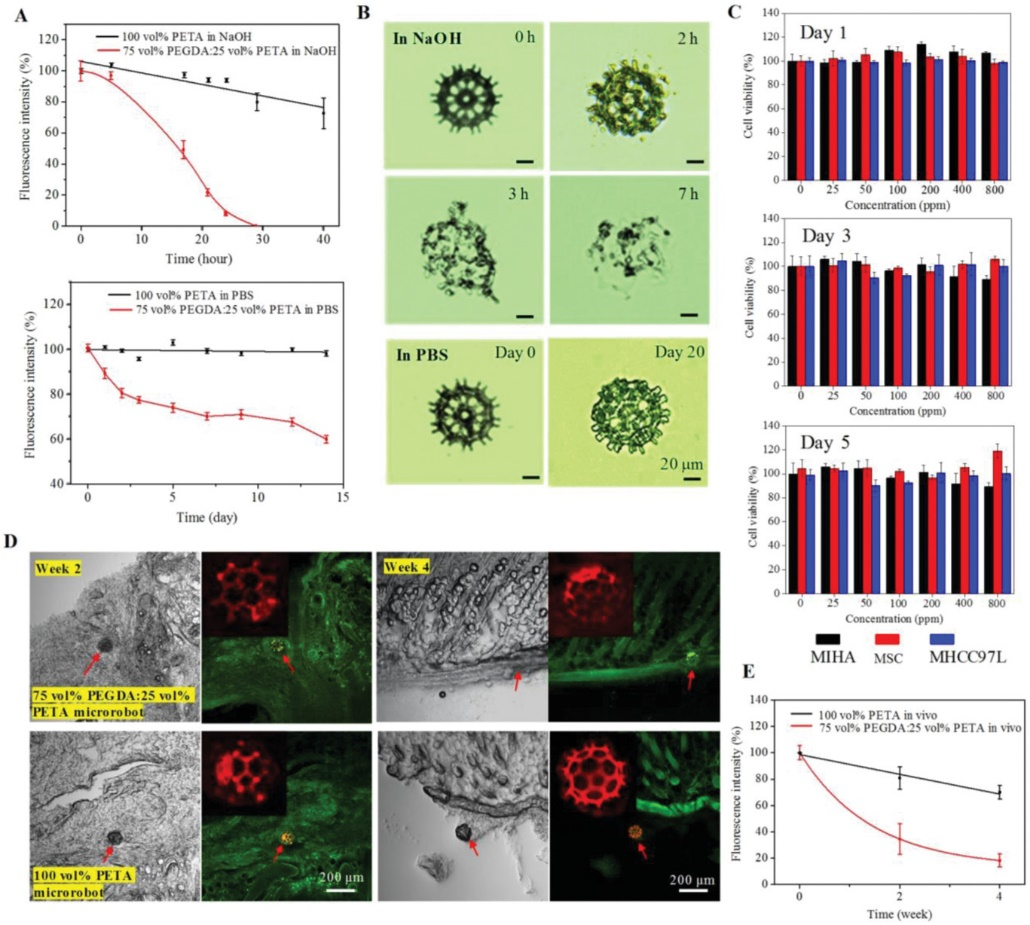
Figure 2: Degradability and cytotoxicity of microrobots. A) Fluorescence decay profile of microrobots made from degradable 75 vol% PEGDA. B) Degradation of microrobots in NaOH solution and PBS. C) MTT assays of three cell types cultured in degradation products at different concentrations for 5 days. The three cell types include therapeutic cells (hiPSC+MSC+GPx3), normal cell line (hepatocyte MIHA), and cancer cell line (hepatocellular carcinoma MHCC97L). D) Confocal scans of histological sections of skin tissues implanted with degradable 75 vol% PEGDA: 25 vol% PETA microrobots (above) and non-degradable 100 vol% PETA microrobots (below). Microrobots appear red or orange, and tissues appear green. E) Fluorescence decay profile of subcutaneously implanted microrobots.
The local tissue environment, enzyme oxidation, and macrophage activity may affect the degradation of the hydrogel. In vivo tests of microrobot degradability were conducted in the subcutaneous tissues of nude mice to provide a realistic environment for studying microrobot degradation. The researchers implanted degradable 75 vol% PEGDA: 25 vol% PETA microrobots and non-degradable 100 vol% PETA microrobots subcutaneously on either side of each mouse. Confocal scan images showed that the microrobots were not washed away. Considering that the degradation products of the microrobots will remain in the body for some time, the researchers subsequently conducted feasibility tests to check the biocompatibility of the fully degraded products. Even at ultra-high concentrations of degradation products, the viability of all cell types exceeded 80%. The cell morphology and proliferation in the experimental group were similar to those in the control group. These results indicate that the biocompatibility of the microrobots was maintained during the 5-day testing period.
2. Cell Release from Microrobots
After the cell-loaded microrobots reach the target site, the release of cells from the microrobots determines the effectiveness of cell therapy. The protein-repellent properties of PEGDA enable cells to migrate actively from the microrobots. The degradation and expansion of the microrobots will aid in the passive separation of cells from the surface of the microrobots. This study first evaluated the cell release capability on glass substrates. Four cells separated from the microrobots on Day 1 were observed to release and expand into a colony after 5 days of culture. The second experiment was conducted using hepatocyte cell lines (MIHA) on a matrix to simulate the liver tissue environment. During 3D culture, a large number of GPSC+MSC+GPx3-labeled GSPs were found to migrate from the microrobots to the MIHA region. Subsequently, experiments were performed on a microfluidic chip to simulate transcutaneous migration of cells transported from within blood vessels. Approximately 200 microrobots loaded with hiPSC+MSC+GPx3 were transported along vessel-like microchannels to docking areas. These cells were released from the microrobots and migrated through connected gaps into the Huh7 cell tissue chamber (hepatocellular carcinoma cell line). The above results confirm that hiPSC+MSC+GPx3 can spontaneously release from the designed microrobots. The design also assessed the inhibitory effects of released cells on Huh7 cells. Another culture with the same number of Huh7 cells was performed alongside the control group. After 3D proliferation, the density of cells in the treatment group was significantly lower than that in the control group. This finding indicates that the released cells can slow the growth of Huh7 cells by simulating the endothelial layer.
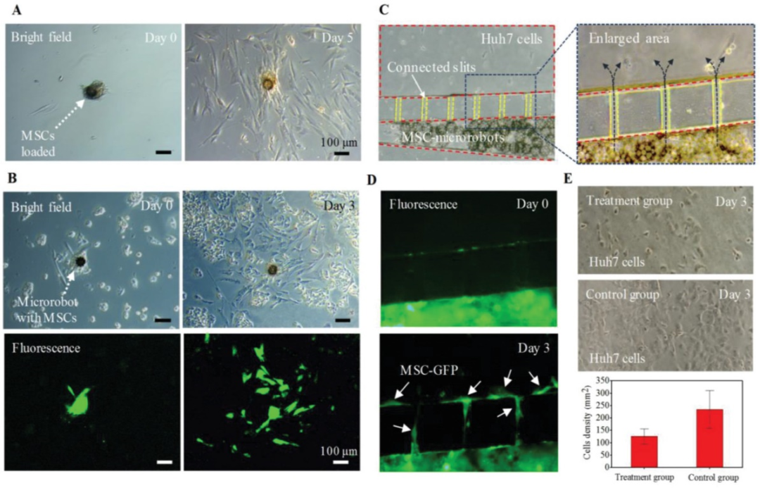
Figure 3: HiPSC+MSC+GPx3 (GFP) released from microrobots to external target sites. A) MSC released from microrobots onto glass substrates. B) MSC labeled with GPC released from microrobots onto matrices cultured with MIHA cells, proliferating during 3D culture. C) MSC released from microrobots in the microfluidic chip for co-culture with Huh7 cells. D) Fluorescence images of MSC released from microrobots and migrated to the Huh7 cell chamber. E) Phase contrast images and statistical results for Huh7 cells.
3. PA Imaging Feedback-Guided Automatic Navigation of Microrobots
To achieve precise delivery of therapeutic cells carried by microrobots, two challenging issues must be addressed. First, the cell-loaded microrobots should be capable of automatically navigating to the desired site with precision. Second, an in vivo imaging technology should be developed to track the microrobots in deep tissues (clinical depth reaching 2 cm). In vitro experiments were first conducted to guide cell-loaded microrobots through “Y” shaped microchannels using a self-made electromagnetic coil system. A precision fluid pump provided controllable flow rates for the liquids in the microfluidic channels. The magnetic control performance of the microrobots exhibited characteristics of three biological fluids in a static environment for the first time. The experimental section detailed the control of microrobots along paths a=b and b=c in a flowing environment. In the experiments, a vision-based proportional-integral-derivative (PID) controller was used to determine the input current for the coils. The cell-loaded microrobots were automatically navigated in an in vitro environment of nude mice. The microrobots loaded with cells were transferred to gastric tissues collected from nude mice (male, 6-8 weeks) and moved along closed-loop paths a=b, b=c, c=d, and d+a. To overcome resistance caused by viscosity and other large viscous molecules in the environment, the microrobots could only move when the magnetic field gradient reached 8.7 T/m, resulting in near-zero positional error in each path, indicating that the microrobots can navigate precisely to the desired location in an in vitro environment.
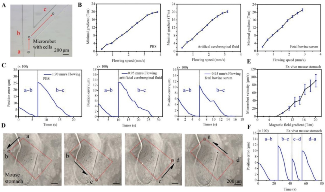
Figure 4: Navigation control of magnet-driven microrobots, both in vitro and in vivo loaded with cells.
To facilitate in vivo experiments, real-time imaging of microrobots with depths ranging from a few millimeters to centimeters is required. We designed the microrobots and customized PA tomography (PAT) for in vivo imaging of microrobots. The microrobots were designed using optical absorption materials to ensure high optical absorption coefficients and high contrast with hemoglobin molecules in the visible spectrum. By tuning the optical excitation wavelength, the contrast of microrobots in blood was optimized while quantifying both blood and microrobots.
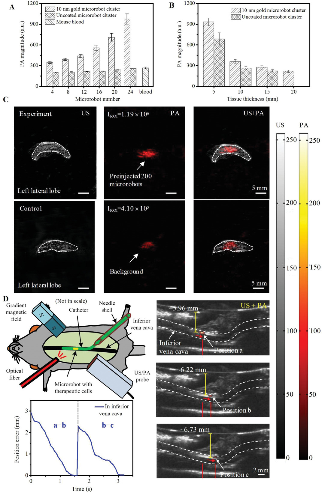
Figure 5: PA imaging of microrobots. A) PA sizes of microrobot clusters with different numbers of microrobots. B) Clustering of PA sizes of microrobots against tissue thickness. C) US and PA images of microrobots. The imaging depth of the microrobots was 6 mm. D) Image-guided navigation of cell-loaded microrobots.
Unlike recently reported work, the researchers conducted their work in blood vessels, where the strong background PA signal from blood complicates the vascular environment. The precise navigation of several microrobots indicates that this therapy is effective, yet challenges remain in real-time imaging in deep tissues. To our knowledge, this is the first time that cell-loaded microrobots have been magnetically driven and optoelectronically navigated within blood vessels in vivo.
4. Preclinical Testing of MSC-Loaded Microrobots for Liver Tumor Treatment
To verify the therapeutic effects of the developed microrobots for cell delivery, experiments were conducted on nude mice implanted with orthotopic liver tumors, delivering hiPSC+MSC+GPx3 via microrobots. The carried hiPSC+MSC+GPx3 was designed to produce GPx3 protein through plasmid design, inhibiting epithelial-mesenchymal transition via the extracellular signal-regulated kinase + nuclear factor interaction protein signaling pathway, thereby suppressing tumor growth and aggressiveness. Cells without GPx3 overexpression could not exert inhibitory effects on cancer cells. The nude mice were divided into three groups: a treatment group receiving MSC from microrobots, a non-treatment group with microrobots carrying no MSC, and a control group. In the treatment group, each mouse implanted with a tumor received 200 MSC-loaded microrobots. In the non-treatment group, the same number of microrobots without MSC was injected into each mouse. In the control group, only PBS was injected into each mouse. The in vivo cell release capability of MSC microrobots was evaluated in the treatment group. The treatment group exhibited significantly more severe tumor cell apoptosis compared to the control group. All these results indicate that microrobots are effective in delivering cells to tumor sites for cancer therapy.
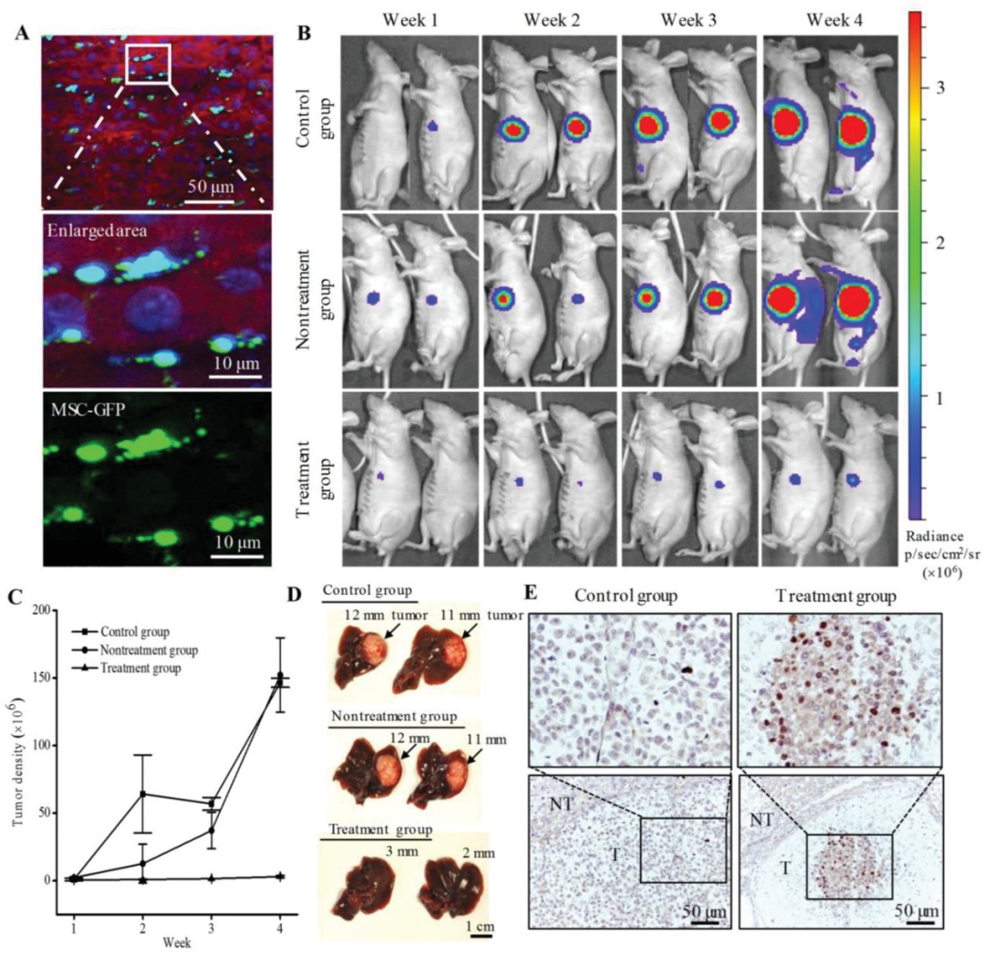
Figure 6: In vivo experiments delivering hiPSC+MSC+GPx3 via microrobots for liver tumor treatment.
Degradable microrobots are designed to meet the requirements of degradability, structural integrity, and magnetic driving capability for in vivo applications. Secondly, real-time PA imaging technology is used to track microrobots in blood vessels. Due to high temporal and spatial resolution, high molecular contrast, and deep penetration, PA imaging can locate and navigate small numbers of microrobots in deep vascular tissues. Thirdly, preclinical tests on live animals demonstrate that microrobots can carry and deliver therapeutic cells to tumor sites for cancer treatment. This study provides a reference for precise treatment methods for diseases such as cancer through wireless and minimally invasive approaches.
— Long press the QR code to read the original English text —

Paper link:
https://doi.org/10.1002/smll.201906908
Experts and scholars are welcome to submit manuscripts (research results, cutting-edge technologies, academic exchanges, etc.). Submission email: [email protected] Top 10 Most Viewed Articles
-
Submission Guidelines for Cross-Disciplinary Medical Engineering V1.0
-
Review Highlights: Progress of Hyaluronic Acid in Tissue Engineering Applications
-
Biomaterials: GelMA/HAMA Dual-Component Hydrogels Loaded with Adipose Stem Cells Promote Skin Wound Vascularization
-
Light-Curing GelMA Hydrogel (EFL-GM Series)
-
Light-Curing Hyaluronic Acid Hydrogel (EFL-HAMA Series)
-
Custom High-Precision 3D Printed Hydrogel Bio-Scaffolds
-
Custom High-Precision 3D Printed Polymer Bio-Scaffolds
-
Light-Curing Bio 3D Printing Printability Evaluation Standards and Printing Process Models
-
Lightweight 3D Cell Culture Solutions Based on Light-Curing Hydrogels
-
EFL’s Third Generation Multifunctional Extrusion Bio 3D Printer Coming Soon!

Tap “Looking” to share