 Introduction
Can a “bridge” be built at the site of a spinal cord injury to reconnect nerve signals? A team from the University of Minnesota published groundbreaking research in Advanced Healthcare Materials: using 3D printed scaffolds to guide stem cells to successfully differentiate into spinal organoids in vivo, which, after transplantation, successfully “reconnected” severed nerve signals in rats, significantly restoring motor function. This technology provides a new strategy for spinal cord injury repair.
01Research Background
Spinal cord injury (SCI) poses a significant health burden globally, with current clinical methods only able to improve quality of life, lacking effective therapies to promote nerve regeneration. Although region-specific spinal neural progenitor cell transplantation can reconstruct host neural connections, it faces challenges such as imprecise cell localization and unclear mechanisms of action. Traditional cell injections lead to cell loss due to lack of structural support, while existing 3D printed scaffolds, although providing mechanical guidance, struggle to meet all biological requirements, and their integration with organ-like structures is still in the exploratory stage. Previous clinical trial failures further highlight the necessity of cell-scaffold synergistic therapy.
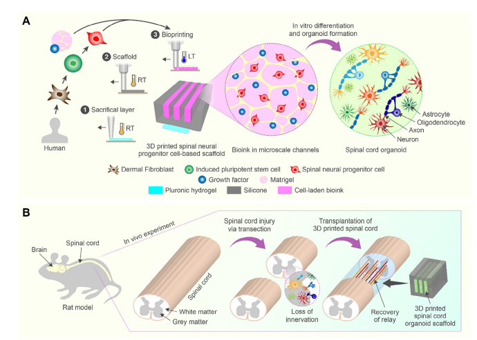
This study proposes a groundbreaking solution: constructing silicone scaffolds with microchannels through multi-material 3D printing technology, combined with clinically-grade region-specific sNPCs derived from human iPSCs, to directionally print cells within the channels and guide their differentiation into spinal-specific neurons, ultimately forming a biomimetic spinal organoid scaffold. This design simulates the spinal gray matter structure through microchannels, achieving axonal directional extension and neural network assembly, laying the foundation for establishing a neural relay system across the injury site.
02Research Overview
Based on the design of functionalized conductive polymers, the research team developed a functionalized polyaniline-based sequential adhesive hydrogel patch. It enables synchronized mechanical physiological monitoring and electrical coupling treatment of the heart, firmly adhering to the heart’s surface to monitor its mechanical movements and electrical activities.
1. 3D Printed Scaffold Design and Organoid Formation Mechanism
This study constructed a silicone scaffold with a microchannel structure (200μm wide) using multi-material extrusion 3D printing technology. Region-specific spinal neural progenitor cells (sNPCs) derived from human iPSCs were combined with Matrigel bioink and precisely printed into the channels. The mechanical guidance of the scaffold prompted sNPCs to directionally extend axons (SMI312+), and within 40 days, they differentiated into V0/V1/V2a spinal interneurons (Exx1+/FOXP2+/Chx10+), forming a layered organoid structure. In vitro culture demonstrated that 78.35% of the cells differentiated into mature neurons (MAP2+), and the neural network could stably maintain for at least one year. RNA sequencing further revealed that the 3D environment significantly upregulated spinal regionalization genes (such as HOXB4/HOXA5) and promoted functional neuronal maturation.
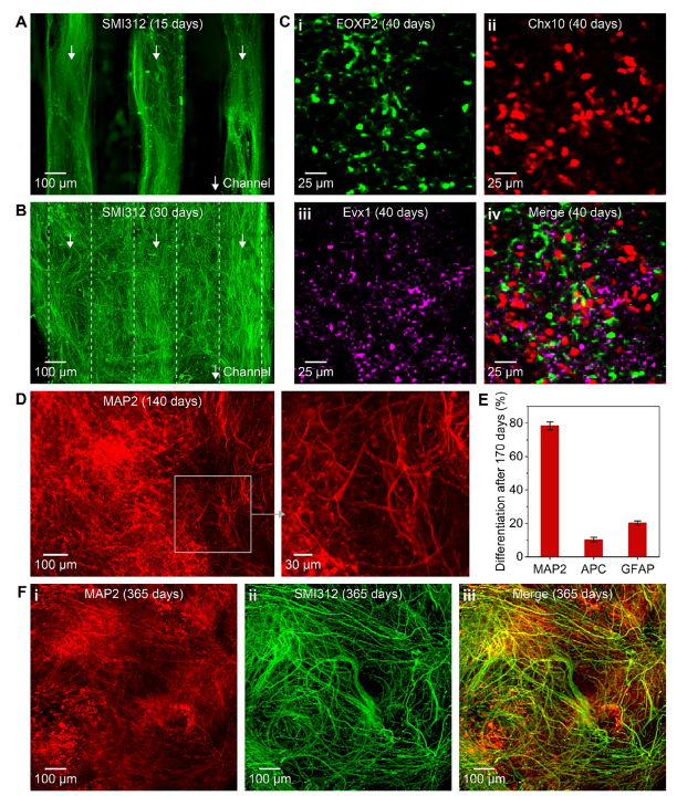 Figure 1: Immunohistochemical results of 3D printed spinal scaffolds in vitro
(Image from the original text)
2. Core Evidence of In Vivo Transplantation Promoting Functional Recovery
The organoid scaffold cultured for 40 days was transplanted into the complete transverse injury area of the rat spinal cord, significantly improving motor function: BBB scores increased from 4.8 at week 3 to 8.4 at week 12 (control group ≤3.6), while the amplitude of motor-evoked potentials (MEP) increased 2.6 times (2.18 mV vs. 0.83 mV). Histological analysis confirmed that the transplanted cells (SC121+) extended axons (NF200+) along the scaffold channels towards the host tissue at both ends, bridging the injury site. Quantitatively, 63.1% of the cells differentiated into neurons, and 20.2% into oligodendrocytes, with synapse formation detected at the host-graft interface, indicating the establishment of a functional neural relay system.
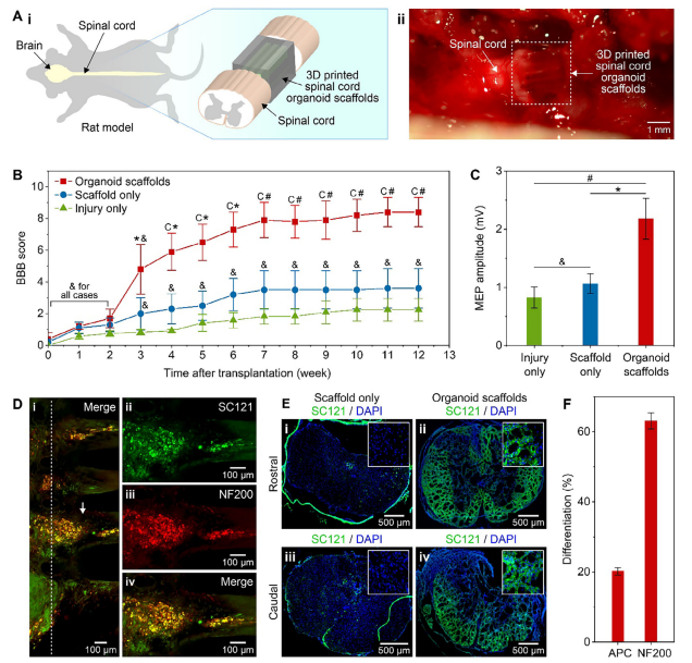 Figure 2: Functional recovery of rats after transplantation of 3D printed organoid scaffolds
(Image from the original text)
3. Technical Breakthrough and Translational Value
This study integrates 3D printed microchannel scaffolds, region-specific sNPCs, and organoid assembly technology for the first time, addressing the challenge of disordered integration of cells in spinal cord injury repair. The directional guidance of microchannels allows axons to precisely cross the injury site, while the multi-type neurons differentiated from sNPCs successfully reconstruct the host neural circuits. The dual scaffold dorsal-ventral assembly strategy further optimizes the efficiency of neural signal relay. This platform provides a transformative therapeutic strategy for chronic spinal cord injury, combining structural support (silicone scaffold) and biological activity (organoids), laying the foundation for clinical translation.
Figure 3: Experimental schematic diagram
(Image from the original text)
From scaffold design, organoid maturation to animal validation, this study achieves precise reconstruction of neural structures and functional recovery after spinal cord injury through interdisciplinary technological integration, providing a new paradigm for regenerative medicine.
03Research Significance
This study successfully constructs a neural relay system in a rat model of complete spinal cord transection by guiding human iPSC-derived spinal neural progenitor cells to form region-specific organoids using 3D printed microchannel scaffolds, significantly promoting functional recovery. This strategy provides a new platform technology for regenerative treatment of spinal cord injuries, combining structural support and biological activity, with significant clinical translation potential.
References:
Guebum Han, Nicolas S. Lavoie, Nandadevi Patil, Olivia G. Korenfeld, Hyunjun Kim, Manuel Esguerra, Daeha Joung, Michael C. McAlpine, Ann M. Parr. 3D‐Printed Scaffolds Promote Enhanced Spinal Organoid Formation for Use in Spinal Cord Injury. Advanced Healthcare Materials, 2025; DOI: 10.1002/adhm.202404817
END
Integrating resources from industry, academia, and research to disseminate cutting-edge technology information. The BME Rehabilitation Engineering Branch’s public account aims to become a professional, authoritative, and high-level communication platform in the field of rehabilitation engineering.
We warmly welcome experts and scholars to contribute!
Submission email:[email protected]