Journal: Lab on a Chip (IF=5.4)
Publication Date: July 14, 2025
Original Link: https://doi.org/10.1039/d5lc00181a
Researchers from the University of Bern, Switzerland, proposed a microfabrication method that combines 3D printing technology with standard fluorescence microscopy, achieving maskless lithography through Digital Micromirror Device (DMD). This method enables micron-level precision micro-manufacturing without the need for a cleanroom. The approach significantly reduces material and time costs and is suitable for various biological applications, including cell morphology regulation, microfluidic chip fabrication, and long-term in vivo imaging, providing new tools for biomedical research and promoting the widespread application of micro-manufacturing technology in the biomedical field. The research results were published in the journal Lab on a Chip under the title “Teach your microscope how to print: low-cost and rapid-iteration microfabrication for biology”.

✦
✦
Background Knowledge
✦
🔵 Traditional microfabrication techniques such as photolithography and soft lithography can achieve high-precision microstructure manufacturing but rely on cleanrooms, expensive equipment, and toxic materials, with long iteration cycles that limit their widespread application in biological research.
🔵 In recent years, the development of 3D printing technology has provided new ideas for rapid prototyping, but its precision often fails to meet the demands for micron-scale structures in biological research.
✦
✦
Research Methodology
✦
🔵 Equipment Modification: Utilizing standard fluorescence microscopy equipped with a Digital Micromirror Device (DMD) and UV light source, the DMD’s pattern projection is computer-controlled to project UV light onto a glass slide coated with photosensitive resin, curing the resin in the exposed areas while washing away the unexposed areas to form the desired microstructure mold.
🔵 Material Selection: Using consumer-grade 3D printing resin instead of traditional SU-8 photoresist and ordinary glass slides instead of silicon wafers, reducing material costs and operational complexity.
🔵 Process Flow: Includes steps such as pre-treatment of the glass slide (coating with TMSPMA to enhance resin adhesion), spin-coating of photosensitive resin, UV light projection exposure, washing of uncured resin, post-curing of the mold, and PDMS replication.
🔵 Automated Control: Achieved through μManager software combined with Python scripts, enabling automated control of the microscope, improving printing efficiency and precision.
✦
✦
Key Conclusions
✦
🔵 This microfabrication method has advantages such as low cost, rapid iteration, ease of operation, and strong compatibility, meeting the demands for micron-level precision structures in biological research.
🔵 The method has broad application prospects in cell biology, tissue engineering, and microfluidic chip fabrication, promising to drive innovation and development in biological research.
✦
✦
Image Analysis
✦
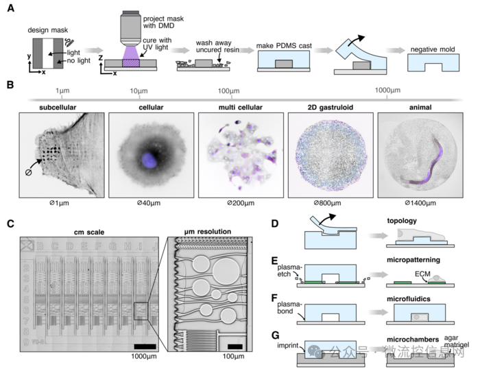
Figure 1 illustrates the overall process of maskless lithography microfabrication based on microscopy. From mask design to final PDMS replication, the entire process utilizes standard fluorescence microscopy and DMD systems to manufacture microstructures through UV light projection, enabling the rapid and low-cost production of micron-level precision structures suitable for biological research, covering various biological scales from subcellular to tissue.
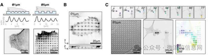
Figure 2 shows the impact of microfabrication technology on cell behavior. Cells form enriched actin structures on surfaces with micron-scale pits or pillars, indicating that cells can sense and respond to the microtopography of the surface. This technology can effectively regulate cell morphology and behavior, providing a powerful tool for cell biology research.
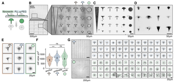
Figure 3 demonstrates the application of micro-patterning technology in cell morphology regulation. By designing micro-patterns of different shapes, cells can be guided to form specific morphologies, such as anchor-shaped, umbrella-shaped, and circular. The attachment of fluorescently labeled cells on different micro-patterns and the quantitative analysis of cell morphology indicate that this technology can precisely control the growth environment of cells, providing new means to study the relationship between cell morphology and function.
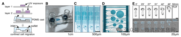
Figure 4 showcases microfluidic chips manufactured using this microfabrication technology. It details the manufacturing process of the chip, including the stacking of multilayer structures, formation of channels, and connection to external devices. The results of cell migration experiments in the microfluidic chip demonstrate its effectiveness in studying cell migration behavior, providing support for research on cell behavior in complex microenvironments.
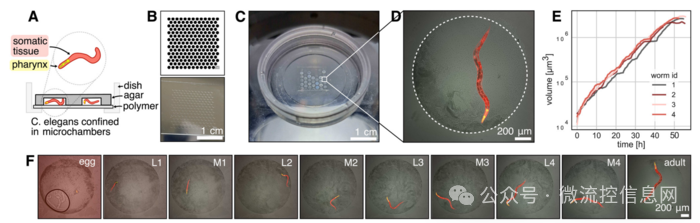
Figure 5 illustrates the agarose microchamber manufactured using microfabrication technology for long-term tracking of Caenorhabditis elegans growth. The manufacturing process of the microchamber, the behavior of the nematodes within it, and the results of long-term imaging indicate that this technology can provide a stable microenvironment for long-term observation of live organisms, holding significant application value for studying the developmental processes of organisms.
✦
✦
Summary and Outlook
✦
In this article, an innovative microfabrication technology is introduced, which modifies a standard fluorescence microscope and combines it with 3D printing technology to achieve low-cost, rapid-iteration microstructure manufacturing. This technology shows broad application prospects in cell biology, microfluidic chip fabrication, and in vivo imaging, promising to drive innovation and development in biological research.
Future research can further optimize the precision and efficiency of this technology, developing more microstructure designs and manufacturing methods suitable for biological research. Additionally, integrating automation technology and artificial intelligence is expected to realize a more intelligent microfabrication process, providing stronger technical support for biological research.