
Real-time biochemical monitoring is crucial in personalized medicine and disease diagnosis. Fiber electrochemical sensors have become ideal carriers for wearable devices due to their miniaturization and biocompatibility advantages. However, traditional surface functionalization preparation strategies lead to two major bottlenecks: first, the active materials are prone to delamination from the high-curvature fiber surface, resulting in mechanical degradation in dynamic biological environments (such as tissue deformation and immersion in body fluids), leading to monitoring failure; second, the uniformity of the coating is difficult to control, with batch performance differences reaching up to 80%, hindering large-scale applications.
Fudan UniversityAcademician Peng Huisheng and Associate Professor Sun Xuemei and their team have developed a universal co-extrusion strategy that achieves continuous preparation of flexible fiber electrochemical sensors by co-extruding active materials with the conductive polymer PEDOT:PSS suspension.This technology allows for the uniform embedding of active materials within an interpenetrating conductive polymer network, forming a stable interfacial structure. The resulting sensors exhibit excellent stability and consistency (performance deviation < 5%) in complex biological environments, successfully applied for dynamic monitoring of hydrogen peroxide (H₂O₂) in subcutaneous mice and continuous tracking of ascorbic acid (AA) in the brain for up to 14 days.
Figure 1 reveals the innovative structure of the co-extruded fiber sensor (EFS). The active materials of traditional coated sensors (Figure 1a-i) are prone to falling off due to bending or friction, while EFS (Figure 1a-ii) addresses the structural instability issue at its source through the uniform integration of active materials with the polymer network. The preparation process (Figure 1b) shows: mixing active materials (such as MnO₂ nanoparticles), PEDOT:PSS, dimethyl sulfoxide (DMSO), and dodecylbenzenesulfonic acid (DBSA) and extruding them into a silicone tube, followed by spontaneous gelation, washing, and drying, resulting in continuous fibers with a diameter of 42.31±0.52 μm (Figure 1c). The diameter and bending stiffness deviation of 50 groups of EFS are both < 5% (Figure 1d), and SEM images (Figure 1f-g) and manganese element distribution maps (Figure 1h) confirm the uniform dispersion of active materials in the fiber cross-section, constructing a stable porous electrochemical active interface.
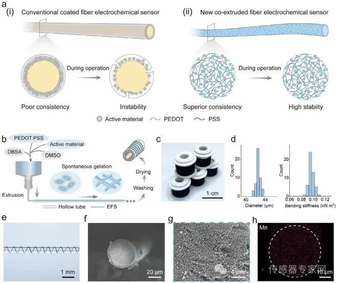
Figure 1: Preparation and structure of the co-extruded fiber sensor (EFS) a) Schematic diagram of traditional coated fiber sensors (i) and EFS (ii). EFS uniformly embeds active materials into the conductive polymer network, exhibiting excellent stability in complex environments. b) EFS preparation process: mixing the DBSA-containing PEDOT:PSS solution with DMSO and active materials, then extruding into a silicone tube, followed by spontaneous gelation, washing, and drying. c) Photo of the continuously produced EFS spool. d) Diameter (left) and bending stiffness (right) distribution of 50 EFS-H₂O₂. e) Flexible display of EFS wrapped around the surface of a capillary. f,g) Scanning electron microscope images of the EFS-H₂O₂ cross-section. h) Energy spectrum map of the uniform distribution of manganese elements in the EFS-H₂O₂ cross-section.
Figure 2 validates the superior performance of EFS. Taking the hydrogen peroxide sensor (EFS-H₂O₂) as an example, its electrochemical active area (10.03 cm²/cm²) far exceeds that of coated sensors (1.24 cm²/cm²) (Figure 2a). At a voltage of -0.4 V, the response current to 1 mM H₂O₂ reaches 87.57 μA/cm², with sensitivity improved nearly 10 times (Figure 2b-c). The sensitivity deviation of 50 groups of EFS is only 4.9% (Figure 2e), far superior to the 80% of coated sensors (Figure 2f). After 1000 bending cycles (radius 2.5 mm) and friction cycles, the sensitivity decay is < 10% (Figure 2g-j), and there is no significant performance decline after soaking in PBS for 14 days (Figure 2k-l), highlighting its mechanical and environmental stability.
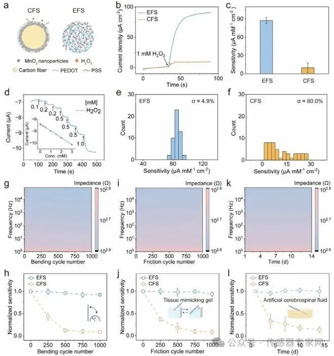
Figure 2: Electrochemical sensing performance, consistency, and stability of EFS a) Comparison of the structure of coated carbon fiber H₂O₂ sensors (CFS-H₂O₂) and EFS-H₂O₂. b) Current response of CFS-H₂O₂ and EFS-H₂O₂ to the same concentration of H₂O₂. c) Comparison of sensitivity between the two sensors (n=50). d) Step current response of EFS-H₂O₂ (inset shows current variation with H₂O₂ concentration). e,f) Sensitivity distribution of 50 EFS (e) and CFS (f). g,h) Impedance (g) and sensitivity variation (h) of EFS-H₂O₂ after 1000 bends. i,j) Impedance (i) and sensitivity variation (j) after 1000 friction cycles. k,l) Impedance (k) and sensitivity variation (l) during 14 days of PBS soaking.
Figure 3 demonstrates the universality of the technology. By changing the active materials, the team successfully prepared ascorbic acid sensors (EFS-AA, Figure 3a) and glucose sensors (EFS-Glucose, Figure 3d). EFS-AA has a linear detection range for AA of 1-600 μM (Figure 3b), selectively excluding neurotransmitter interference (Figure 3c); EFS-Glucose achieves a sensitivity of 198.80 μA/mM/cm² for glucose in 0.1 M NaOH (Figure 3e), effectively resisting interference from ascorbic acid and uric acid after Nafion modification (Figure 3f). Both maintain stable performance under dynamic deformation and long-term immersion (Figure 3g-i).
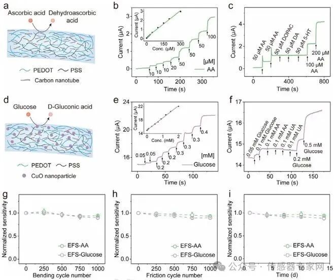
Figure 3: Universality of the co-extrusion strategy a) Schematic diagram of the EFS ascorbic acid sensor (EFS-AA). b) Current response of EFS-AA (inset shows current variation with AA concentration). c) Interference resistance performance test of EFS-AA. d) Schematic diagram of the EFS glucose sensor (EFS-Glucose). e) Current response of EFS-Glucose (inset shows current variation with glucose concentration). f) Interference resistance performance test of EFS-Glucose. g-i) Sensitivity retention of EFS-AA and EFS-Glucose after bending (g), friction (h), and immersion (i).
Figure 4 confirms the in vivo application potential. After implanting EFS in the subcutaneous tissue of mice for 7 days, there was no inflammatory response in the surrounding tissue (Figure 4a-b), achieving dynamic monitoring of subcutaneous H₂O₂ (Figure 4c), and maintaining stable signals under pressure, bending, and friction disturbances (Figure 4d). More remarkably, the implanted EFS-AA in the brain operated continuously for 14 days, with no decay in the response current amplitude to ascorbic acid (Figure 4e-g), providing a new tool for chronic neurological disease research.
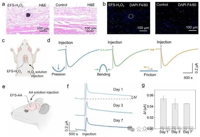
Figure 4: Long-term monitoring of biochemical substances in vivo a,b) H&E staining (a) and immunohistochemical staining (b) of EFS-H₂O₂ implanted subcutaneously in mice for one week, with blue (DAPI) marking cell nuclei and green (F4/80) marking macrophages. c) Schematic diagram of subcutaneous H₂O₂ monitoring. d) Real-time monitoring stability of H₂O₂ under dynamic deformation (left: pressing; middle: bending; right: friction). e) Schematic diagram of bilateral hemisphere monitoring of AA in the brain. f) Real-time monitoring curve of AA within 7 days post-implantation. g) Current response variation of EFS-AA to AA injection after 7 days of implantation.
Future Prospects
This co-extrusion strategy opens a new path for the large-scale production of high-performance fiber electrochemical sensors. In the future, expanding active materials and integrating multiple fibers is expected to achieve multifunctional monitoring with a single fiber, promoting the practical application of wearable medical devices in health management and chronic disease research.
What are your thoughts on this article? Feel free to leave a comment and share!Share and click to see more, spreading the dynamics of the Chinese sensor industry to more people!
Marking the content you care about, according to the recommendation principles of WeChat public accounts, if you have not marked 【Sensor Expert Network】 as a star on the homepage, you will not be able to receive the latest news in the sensor industry in a timely manner!
Source:Frontiers in Polymer ScienceDisclaimer: The content of this article reflects the author’s personal views and does not represent the views or positions of the Sensor Expert Network. For inquiries, please contact: pengzhenlie (WeChat ID). If you have submission or interview requests, please send an email to:[email protected].