1. 3D Microvascularized Tissue Models Based on Collagen Hydrogel – Laser-Induced Cavitation Molding for Creating Channels and CavitiesIntroduction:The team of Professor Frank Niklaus, Anna Herland, and Göran Stemme from the Micro and Nanosystems Division of KTH Royal Institute of Technology published an article titled “3D Microvascularized Tissue Models by Laser-Based Cavitation Molding of Collagen” in ADVANCED MATERIALS. In this study, the authors reported the creation of channels and cavities with diameters ranging from 20 to 60 µm by in situ 3D patterning of collagen hydrogel using femtosecond laser irradiation. During this process, laser irradiation of the hydrogel generates cavitation bubbles that rearrange collagen fibers, resulting in stable microchannels. These 3D channels can form within cell-laden or organoid-laden hydrogels without affecting the activity outside the lumen, and can be perfused with endothelial cell culture and cell-laden media to form artificial microvessels. Therefore, this method enables the realization of organ-on-a-chip models and 3D tissue models with complex microvasculature.— Click Image View Details —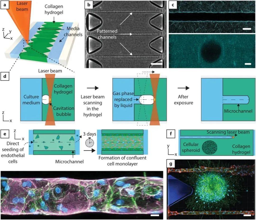
2. Low-Temperature 3D Printed Dual-Delivery Scaffolds Improve Bone Regeneration and Enhance Vascularization
Introduction:Recently, Associate Professor Wang Chong from Dongguan University of Technology, together with teams from Youjiang Medical University for Nationalities, led by Tang Yujin and Liu Jia, employed an extrusion-based low-temperature 3D printing technology to prepare customized bone tissue engineering scaffolds with balanced osteoconductivity/osteogenicity and pro-angiogenic properties. The mechanical structure of the dual-delivery scaffold resembles that of human cancellous bone, allowing for continuous peptide release composed of rapid release (AP) and sustained release (OP). The dual-peptide delivery scaffold not only promotes in vitro angiogenesis but also enhances the in vitro osteogenic differentiation of bone marrow mesenchymal stem cells. In vivo experimental results show that the dual-peptide delivery scaffold promotes new bone formation in rat cranial defects and enhances vascular formation in regenerated tissues. The related paper “Cryogenic 3D printing of dual-delivery scaffolds for improved bone regeneration with enhanced vascularization” was published in Bioactive Materials.— Click ImageView Details —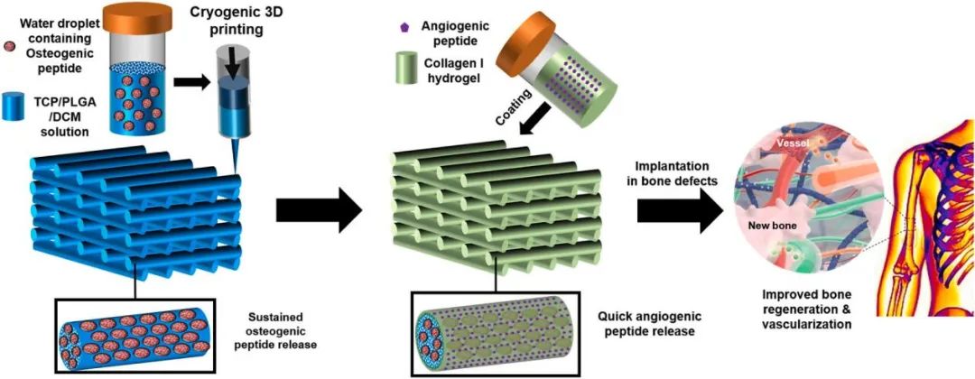
3. Review: Prospects for 3D Bioprinting of Organoids
Introduction:Recently, Preety Rawal, Dinesh M. Tripathi, and Seeram Ramakrishna published a review article titled “Prospects for 3D bioprinting of organoids” in Bio-Design and Manufacturing. The paper focuses on the development and application prospects of organoid biomanufacturing. Three-dimensional (3D) organoids derived from pluripotent or adult tissue stem cells hold great potential in studying developmental and disease mechanisms, as well as various applications in regenerative therapies. However, the lack of precise manufacturing methods for structures and large-sized tissues is a key limitation encountered by current organoid technology. Recent advancements in organoid 3D bioprinting technology attempt to address some of these barriers. This review discusses the use of 3D bioprinting with bioinks and printing methods and highlights recent successful research in stem cell and organoid bioprinting. It summarizes several vascularization strategies for bioprinted organoids, which are crucial for complex tissue construction. To fully realize the translational applications of organoids in disease modeling and regenerative medicine, 3D bioprinting technology should be better utilized in these areas.— Click ImageView Details —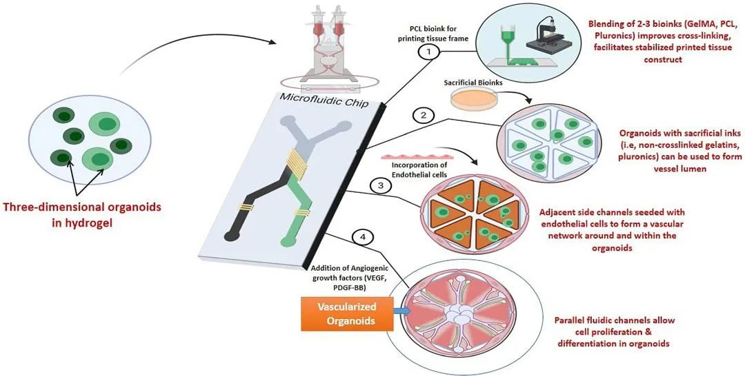
4. 3D Printing of Tissue Engineering Scaffolds: A Focus on Vascular Regeneration
Introduction:Recently, Pengju Wang, Yazhou Sun, and Xiaoquan Shi published an article titled “3D printing of tissue engineering scaffolds: a focus on vascular regeneration” in Bio-Design and Manufacturing. This review paper summarizes the research progress of 3D printed scaffold technology in the field of vascular regeneration. The topic of using 3D printed scaffolds for vascular regeneration has been a research hotspot and is of great importance; however, there is a relative lack of systematic summaries regarding research in this area in existing reports. The article elaborates on five aspects: 1) the significance and importance of tissue engineering scaffolds for vascular regeneration; 2) 3D modeling methods for vascular scaffolds; 3) commonly used 3D printing materials for vascular scaffolds; 4) common 3D printing technologies used in the manufacture of vascular scaffolds; 5) clinical translation of vascular scaffolds. Additionally, considering the advantages of traditional manufacturing techniques, the paper discusses other technologies often involved in the preparation of vascular scaffolds, including casting, electrospinning, and Lego-like construction.— Click ImageView Details —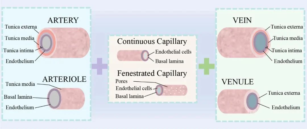
5. In Vivo Study of Conductive 3D Printed PCL/MWCNTs Scaffolds with Electrical Stimulation for Bone Tissue Engineering
Introduction:Recently, Edney P. e Silva, Boyang Huang, and others published an article titled “In vivo study of conductive 3D printed PCL/MWCNTs scaffolds with electrical stimulation for bone tissue engineering” in Bio-Design and Manufacturing. In this study, conductive scaffolds made from biocompatible and biodegradable polycaprolactone (PCL) and multi-walled carbon nanotubes (MWCNT) were produced using an extrusion-based additive manufacturing method to treat large cranial defects in rats. Histological results show that using PCL/MWCNTs scaffolds and electrical stimulation (ES) helps form thicker and increased bone tissue within the bone defect. The use of a high concentration of multi-walled carbon nanotubes (3 wt%) and ES also significantly promotes angiogenesis and mineralization. Additionally, the scaffolds encourage the formation of tartrate-resistant acid phosphatase (TRAP) positive cells, while the addition of MWCNTs appears to inhibit the formation of osteoclasts, though it has limited effects on osteoclast function (expression of RANKL and OPG). The use of ES promotes osteoclast generation and RANKL expression, demonstrating a dominant role in bone remodeling. These results suggest that the combination of 3D printed conductive PCL/MWCNTs scaffolds and ES is a promising strategy for treating critical bone defects and provides clues for establishing optimal protocols for bone tissue engineering using conductive scaffolds and ES.— Click ImageView Details —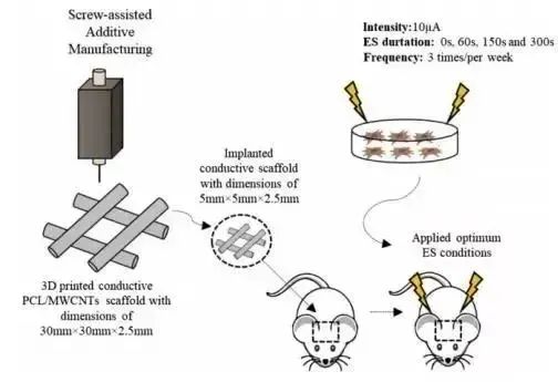
6. 3D Vascularized Liver Tissue Models Based on Multimaterial Printing
Introduction:The research team led by researcher Gu Qi from the Institute of Zoology, Chinese Academy of Sciences, in collaboration with researcher Zheng Xiongfei from the Shenyang Institute of Automation, Chinese Academy of Sciences, and researcher Wang Shu from the Institute of Chemistry, Chinese Academy of Sciences, developed a multimaterial printing method for preparing vascularized tissues using a custom printer, which has great application prospects in drug screening and liver tissue engineering. The related paper “3D Liver Tissue Model with Branched Vascular Networks by Multimaterial Bioprinting” was published in Advanced Healthcare Materials.— Click ImageView Details —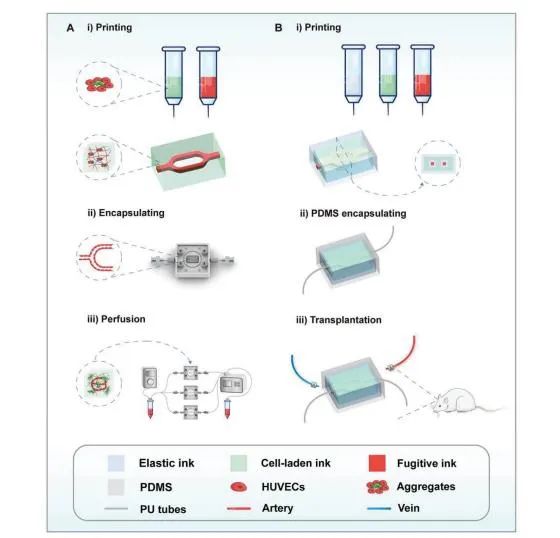
7. Customizable DLP Printed Multimaterial Hydrogel Microfluidic Chips
Introduction:The team led by Amir K. Miri from Rowan University in the USA developed a DLP bioprinter capable of rapidly and simultaneously producing multi-material composite hydrogel microfluidic chips based on polyethylene glycol diacrylate (PEGDA) and methacrylated gelatin (GelMA). These microfluidic chips exhibit good mechanical properties and biocompatibility and can effectively promote microtissue vascularization. This biomanufacturing method can greatly assist in the rapid integration of microtissue models into organ-on-a-chip and high-throughput drug screening platforms. The related paper “Multi-Material Digital Light Processing Bioprinting of Hydrogel-Based Microfluidic Chips” was published in Biofabrication.— Click ImageView Details —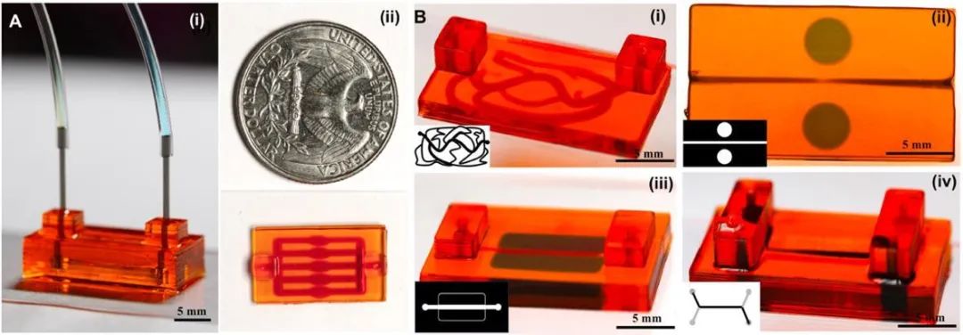
8. Thermoresponsive Hydrogel-Based Bioprinting of Microscale Vascular Networks
Introduction:One of the key challenges in tissue engineering research is to form complex and functional vascular networks in large tissues to maintain oxygen and nutrient delivery and effectively clear waste. Currently, light-assisted processes such as stereolithography, DLP, and selective laser sintering have become the primary tools for fabricating microscale vascular networks (MSV). However, these methods often involve complex preparation processes and require specific photosensitive base materials. Therefore, constructing small-caliber blood vessels and bridging large arteries or veins and capillary networks remains a significant challenge in the field. Recently, the orthopedic team of Wang Jinwu from the Ninth People’s Hospital affiliated to Shanghai Jiao Tong University used thermosensitive hydrogels poly(N-isopropylacrylamide) (PNIPAM) and methacrylated gelatin (GelMA, EFL-GM series) to prepare small-sized MSVs through a sacrificial template method and the contraction effect of thermoresponsive hydrogel scaffolds. At 37°C, the thermosensitive volume contraction of PNIPAM effectively induces the fabrication of smaller-sized MSVs. The related research paper “Fabrication of Thermoresponsive Hydrogel Scaffolds with Engineered Microscale Vasculatures” was published in Advanced Functional Materials.— Click ImageView Details —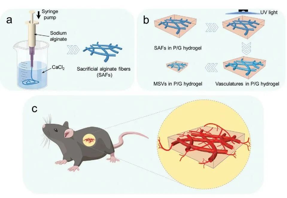
9. Coaxial Printing of Tubular Structures Using F127 Hydrogel
Introduction:Engineered tubular structures made from soft biomaterials have a wide range of applications in biomedicine. The commonly used manufacturing strategy involves casting hydrogel materials around a core, where the diameter of the core determines the lumen diameter of the tube. However, when manufacturing tubes with small lumen diameters (<0.5 mm) and longer lengths (>15 cm), these manufacturing methods are limited. Therefore, coordinating the bioactivity of hydrogel and processability during lumen fabrication remains a challenge. Recently, the team of Alshakim Nelson from the University of Washington in the USA published an article titled “3D printed coaxial nozzles for the extrusion of hydrogel tubes toward modeling vascular endothelium” in Biofabrication, customizing coaxial nozzles using an SLA printer and employing two shear-thinning hydrogels (F-127 and F127-BUM) to coaxially print hydrogel tubes. By designing the three-dimensional model structure of the nozzle, tubes with a lumen diameter of 150 µm can be fabricated.— Click ImageView Details —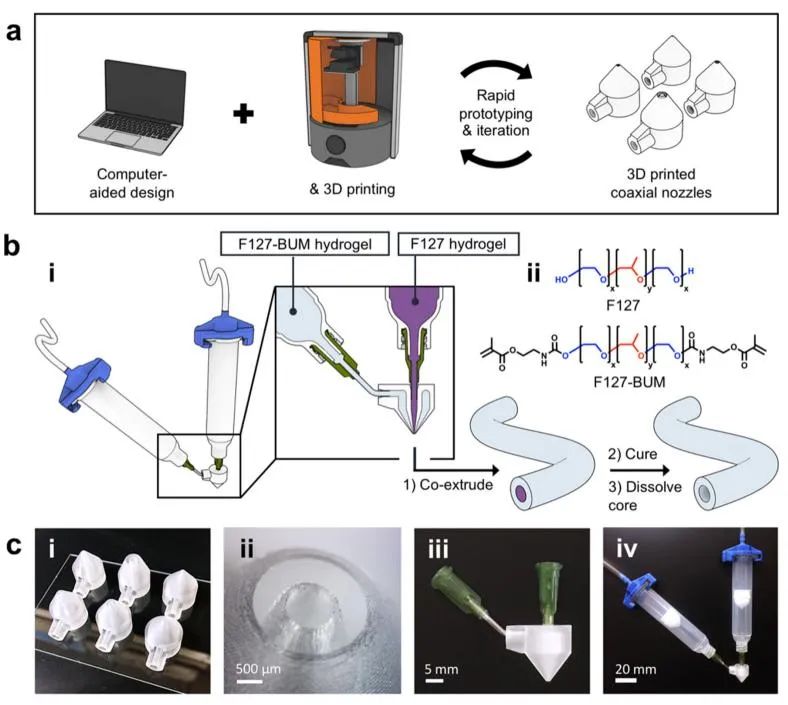
10. Exosome-Eluting Stents for Vascular Repair After Ischemic Injury
Introduction:The team of Ke Cheng from the Joint Department of Biomedical Engineering at the University of North Carolina at Chapel Hill and North Carolina State University published an article titled “Exosome-eluting stents for vascular healing after ischemic injury” in Nature Biomedical Engineering. They developed a bioresponsive exosome-eluting stent (EES) that releases therapeutic exosomes in response to reactive oxygen species (ROS), accelerating vascular healing in rats with renal ischemia-reperfusion injury, and reducing inflammation and smooth muscle cell migration.— Click ImageView Details —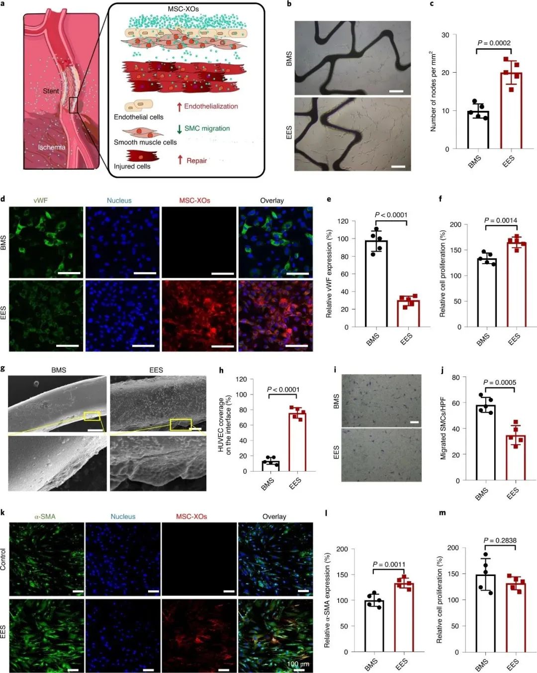
11. Direct 3D Printing of Microvascular Bioink
Introduction:Professor Ying Mei from the Department of Bioengineering at Clemson University in South Carolina published an article titled “Engineering a Chemically Defined Hydrogel Bioink for Direct Bioprinting of Microvasculature” in Biomacromolecules. The team developed an alginate-based hydrogel bioink that can be modified with RGD (a peptide for cell adhesion) and a vascular endothelial growth factor (VEGF) mimetic peptide with a matrix metalloproteinase cleavable linker (MMPQK) for direct fabrication of microvascular tissue structures. This hydrogel can promote vascular morphogenesis and achieve on-demand isolation of migrating cells.— Click ImageView Details —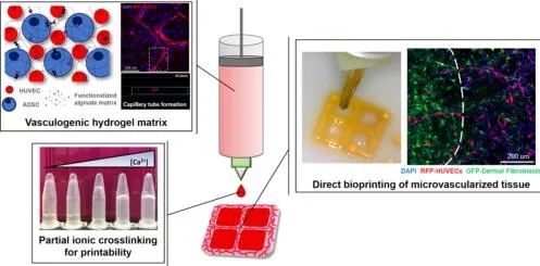
12. AM: Bioprinting of Vascularized Soft Tissues
Introduction:The team of Nasim Annabi at UCLA utilized recombinant human elastin as a highly biocompatible and elastic bioink for the 3D printing of complex soft tissues. Bioprinting of cardiac structures with vascular pedicles was conducted, and their function was evaluated in vitro and in vivo, showing the potential of printing 3D functional cardiac tissues with elastic bioink. The research was published under the title “Human‐Recombinant‐Elastin‐Based Bioinks for 3D Bioprinting of Vascularized Soft Tissues” in Advanced Materials.— Click ImageView Details —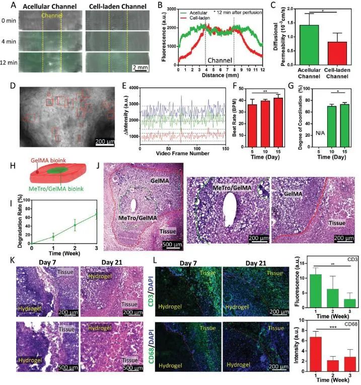
13. AFM: Coaxial Printing of High-Strength Hydrogel Microtubes with Vascular Structures
Introduction:The team of researcher Ruan Changshun from the Shenzhen Institute of Advanced Technology, Chinese Academy of Sciences, in collaboration with researcher Pan Haobo and Professor Liu Wenguang from Tianjin University, successfully constructed a hydrogen-bond reinforced high-strength nano-composite medical hydrogel ink (referred to as CNG printing ink) based on nanoclay/N-acryloyl glycine and gelatin methacrylate (GelMA), combining 3D coaxial printing technology to successfully construct small-caliber microtubes with high toughness, super-stretchability, compressibility, and rapid self-recovery performance, with potential for large-scale production. Furthermore, these microtubes exhibited good biological characteristics, promoting the adhesion, spreading, and endothelialization of endothelial cells, laying the foundation for the application of 3D printed small-caliber microtube scaffolds in tissue regeneration. This research opens up a general and simple method for scaling up the manufacturing of high-strength microtubes, which has immense potential in the regeneration of tubular tissues. The research results were published in Advanced Functional Materials under the title “Coaxial Scale-up Printing of Diameter-tunable Biohybrid Hydrogel Microtubes with High Strength, Perfusability and Endothelialization.”— Click ImageView Details —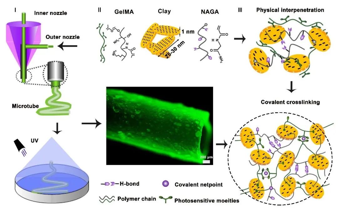
14. Review: Engineering Approaches to Human Vascular Biology from Arteries to Capillaries
Introduction:The team of Professor Gordana Vunjak-Novakovic from Columbia University discusses the design considerations and technologies for engineering millimeter-scale, macro, and microvasculature, further providing examples of recent technologies for engineering multi-scale vascular systems, ultimately identifying key challenges that limit the clinical translation of vascularized tissues and proposing future exploration directions. This review briefly summarizes the structure of vascular trees, their formation during embryogenesis, and their fundamental functions in vivo. It further explores different methods for fabricating vascular systems at various levels, from small-caliber vessels for transplantation to micro and macro-scale flow channels for organ-on-a-chip platforms and engineered tissues. The latest technologies for manufacturing multi-scale vascular systems are further discussed. Throughout the process, a wealth of pioneering papers that influence the development of these subfields will be covered, emphasizing the major progress made in advancing the field over the past few years. Finally, an overview of the current challenges faced by the field and perspectives on future research and manufacturing strategies that may drive the translation of engineered tissues will be provided.— Click ImageView Details —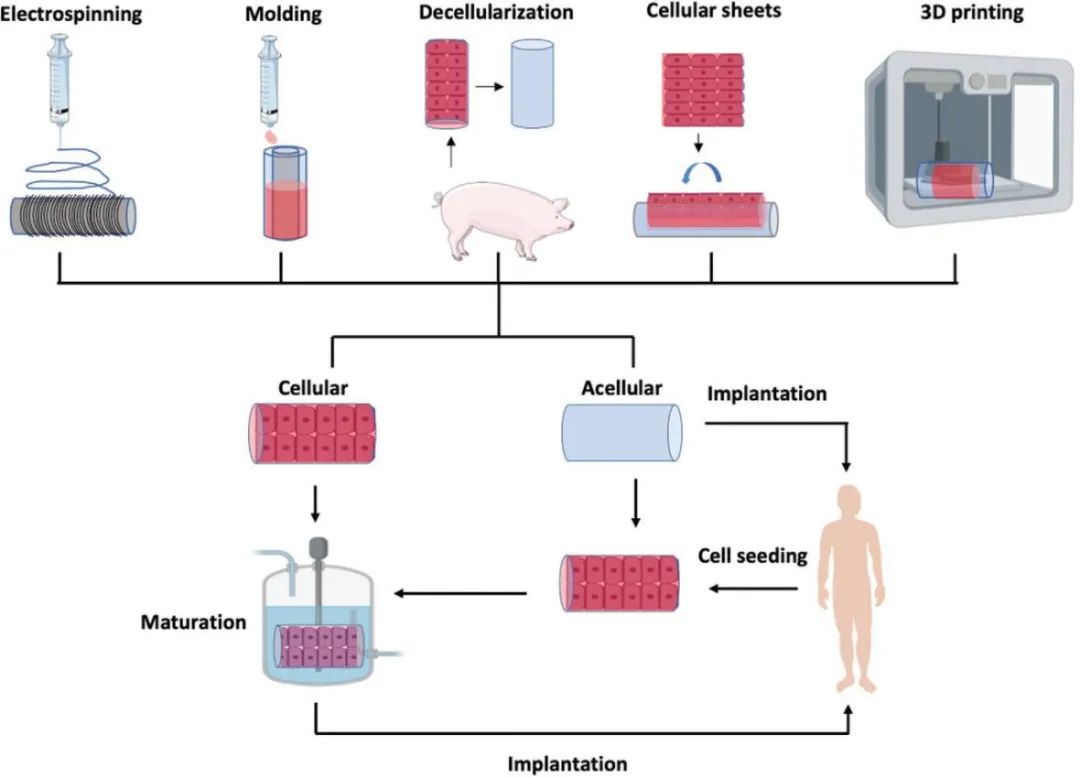
15. AFM: Coaxial 3D Printing of Complex Channel GelMA Hydrogel for Vascular Models and Microfluidic Perfusion Cultures
Introduction:The team of Molly M. Stevens from the Department of Bioengineering at Imperial College London utilizes thermosensitive gelatin-based bioinks as printable sacrificial templates, with photo-crosslinkable GelMA as the filling extracellular matrix template. At 37°C, gelatin spontaneously dissolves to form a continuous vascular network framework. This study achieves the construction of personalized, uniform tubular structure three-dimensional gel networks through a void-free 3D printing method. Compared to other sacrificial ink-based 3D printing methods, this approach addresses the challenges of collapsing 3D gel networks, low viscosity bioinks being difficult to shape, and low efficiency of endothelial cell seeding, and can be used to construct microfluidic chips based on hydrogels with good internal connectivity. The related paper “Void-Free 3D Bioprinting for In Situ Endothelialization and Microfluidic Perfusion” was published in Advanced Functional Materials.— Click ImageView Details —
16. Coaxial Bioprinting Method for Large-Scale Vascularized Tissues
Introduction:Professor He Yong’s team from Zhejiang University proposed a coaxial 3D printing approach for tissue/vascular simultaneous manufacturing by coupling sacrificial printing and coaxial printing processes (Figure 2). By designing a core/shell type GelMA/gelatin ink, the tissue cell-laden ink (outer nozzle) and endothelial cell-laden sacrificial ink (inner nozzle) are simultaneously extruded. During printing, the sacrificial ink supports the flow channels, and during cultivation, the sacrificial ink melts to form unobstructed flow channel networks, while endothelial cells are released from the sacrificial ink and adhere to the channel walls for vascularization, achieving the transformation from flow channel networks to vascularized networks. Based on this method, vascularized large tissue structures (≥1 cm) were printed and cultured for extended periods (≥20 days), successfully applied to the manufacturing of vascularized tumor models and bone tissue. The related paper “Directly Coaxial 3D Bioprinting of Large-scale Vascularized Tissue Constructs” was recently published in Biofabrication.— Click ImageView Details —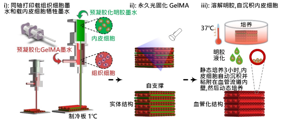
17. Bioprinting of Multiscale Hepatic Lobules within Highly Vascularized Constructs
Introduction:The team of Songwan Jin from Pohang University of Technology in Korea applied a preset extrusion bioprinting technique to create arrays of hepatic lobules, which can simultaneously create heterogeneous, multicellular, and multimaterial structures. The manufactured hepatic lobules include hepatocytes, endothelial cells, and lumens. Endothelial cells surround the hepatocytes, the lobule exterior, and the lumen, ultimately connecting to form a highly vascularized structure. The hepatic lobule structures printed using extrusion bioprinting technology can reach diameters of 1 mm, with a lumen diameter of 150 µm at the center, and the high-density cells in the bioink accelerate tissue formation through interactions between adjacent cells and cell ECM, maintaining structural integrity and cell function, thus enabling the printing of complex heterogeneous hepatic lobule structures into a series of hepatic lobule structure arrays, achieving bioprinting from micro to macro tissues. The related paper “Bioprinting of Multiscaled Hepatic Lobules within a Highly Vascularized Construct” was published in Small.— Click ImageView Details —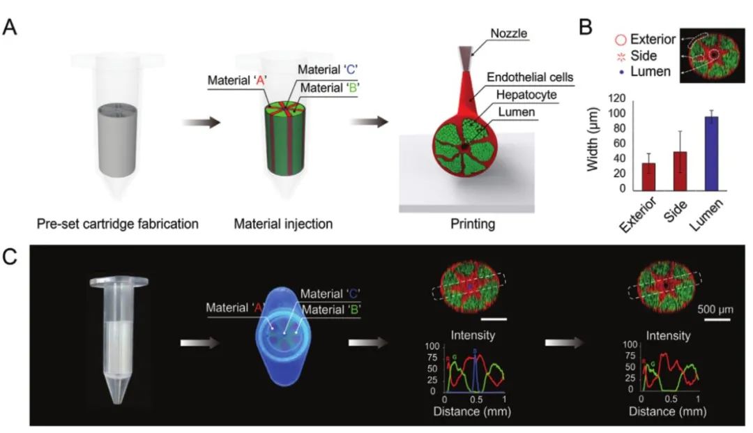
18. Biofabrication of GelMA-Based Vascularized Skin
Introduction:Professor Ali Khademhosseini’s team at the University of California proposed a strategy for constructing three-dimensional skin tissue models: (1) printing GelMA/alginate hydrogels loaded with HUVECs on a polyester porous membrane with a pore size of 0.4 μm, which facilitates the interaction between dermal fibroblasts and endothelial cells and promotes nutrient diffusion (forming internal vascular networks); (2) printing GelMA hydrogels encapsulating human dermal fibroblasts, adjusting matrix stiffness to influence cell growth and function; (3) forming multilayer epidermal gelatin coatings by repeatedly covering the dermal layer with keratinocyte-encapsulated gelatin. This study provides a simple method that uses biomedical and bioengineering techniques to optimize each layer of skin tissue. The constructed 3D skin tissue serves as a multifunctional platform, making the reconstruction of in vitro skin models possible. The related paper “Biofabrication of endothelial cell, dermal fibroblast, and multilayered keratinocyte layers for skin tissue engineering” was published in Biofabrication.— Click ImageView Details —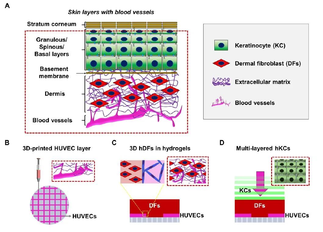
19. 3D Printing of Complete Vascular Networks and Initial Exploration of Tumor-Vascular Interactions
Introduction:Professor He Yong’s team from Zhejiang University proposed a new method for manufacturing multiscale vascular networks on hydrogel materials: by printing multiscale flow channel network templates using a high-precision 3D printer developed by the EFL team, encapsulating hydrogel flow channels using secondary crosslinking principles. This research successfully reconstructed complete blood supply networks (artery-capillary-vein) in vitro, as well as highly branched networks, spiral vessels, and vascular stenosis. A functional vascular network was established in large breast tumor tissues. With the combination of multiscale 3D printing and secondary crosslinking processes, researchers will be able to construct a series of in vitro models for biomedical research, such as studying tumor development processes, exploring tumor-vascular interactions, and screening anti-tumor drugs. The related paper “Construction of multi-scale vascular chips and modeling of the interaction between tumors and blood vessels” has been accepted by the flagship journal Materials Horizons of RSC.— Click ImageView Details —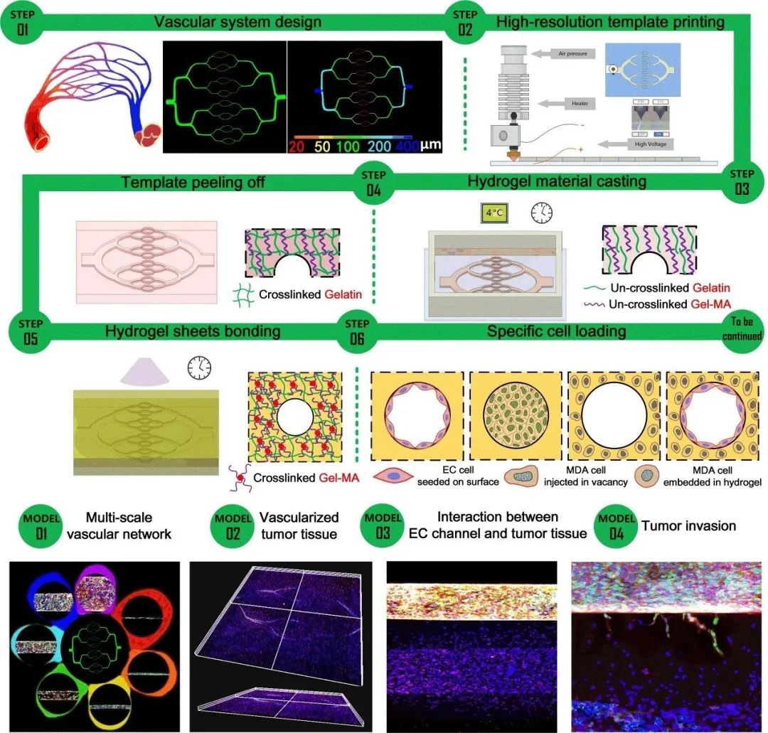
20. TOC-BBL: A Tumor Chip with Bioprinted Blood and Lymphatic Vessels
Introduction:Professor Yu Shrike Zhang’s team at Harvard University designed and constructed a tumor chip platform that simultaneously possesses blood and lymphatic vessels, embedded within a microfluidic bioreactor controlled by a microfluidic system, and achieved the 3D culture of MCF-7 breast cancer cells in a GelMA hydrogel matrix with a dynamic microenvironment. To match in vivo corresponding characteristics, the hollow blood and lymphatic vessels were printed using two different components of bioink, allowing for individual regulation of the permeability of the embedded blood and lymphatic vessels in the hydrogel tumor matrix through optimization of bioink components. Different combinations of blood/lymphatic vessels and cancer cell distributions exhibit completely different diffusion curves for biomolecules and anti-cancer drugs. This in vitro model will be used to study the dynamics and interactions of blood and lymphatic vessels in the tumor microenvironment during the process of anti-tumor drug screening, which is expected to improve the accuracy of drug screening. The related paper “A Tumor-on-a-Chip System with Bioprinted Blood and Lymphatic Vessel Pair” was published in Advanced Functional Materials under WILEY.— Click ImageView Details —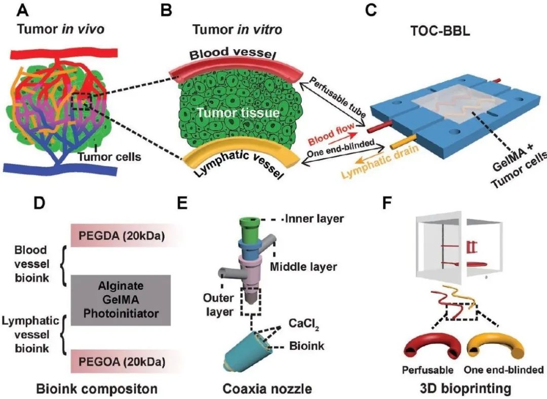
21. Breakthrough in Organ Printing: 3D Printing of High Cell Density and Embedded Vascular-Specific Organ Tissues
Introduction:The research team of Jennifer A. Lewis at the Wyss Institute at Harvard University developed a technique called Sacrificial Writing Into Functional Tissue (SWIFT). In this study, microarray was first used to manufacture hundreds of thousands of OBBs (Organ Building Blocks, dense cellular organoids composed of patient-specific induced pluripotent stem cells). The organ building blocks (OBBs) were then mixed with an ECM solution composed of type I collagen and matrix gel at 0-4°C to form a high cell density living matrix, and finally, perfusable vascular channels were introduced through embedded sacrificial 3D bioprinting. Experimental results show that the SWIFT bioprinting method can rapidly assemble perfusable patient-organ-specific tissues. The related paper “Biomanufacturing of organ-specific tissues with high cellular density and embedded vascular channels” was published in Science Advances.— Click ImageView Details —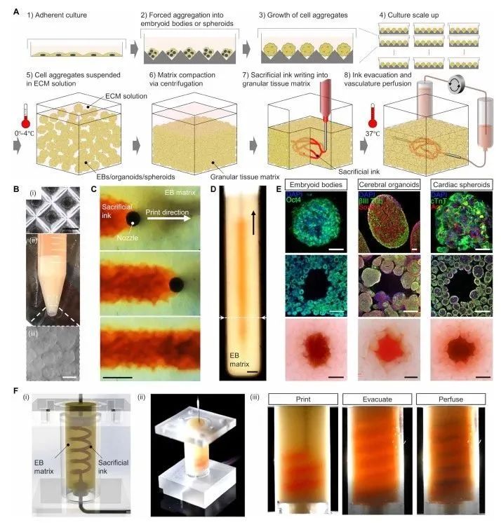
22. Electrospinning + Near-Field Direct Writing to Print Small Diameter Vascular Scaffolds
Introduction:Debby Gawlitta from Utrecht University and Jürgen Groll from the University of Würzburg developed a hybrid manufacturing method combining solution electrospinning with melt near-field direct writing to fabricate a dual-layer composite vascular scaffold that simulates tissue structure, including an inner layer of randomly oriented dense fiber mesh and an outer layer of directionally controllable fibers. This scaffold can induce cells to form a continuous monolayer of endothelial cells and a directionally oriented smooth muscle cell layer, thus promoting tissue-specific cell differentiation. Moreover, this method does not require additional soluble factors or bioactive materials on the scaffold surface, indicating that the structural design of the heterotypic scaffold can induce cell growth and differentiation. This composite structure of the heterotypic scaffold resembles the structure of native vascular intima and adventitia, with the inner electrospun layer having a small diameter and low cell permeability, which can promote the formation of endothelial cell layers, and its future potential can utilize heterotypic designs to achieve modulation of immune responses, steering it towards regeneration. The related paper “Heterotypic Scaffold Design Orchestrates Primary Cell Organization and Phenotypes in Cocultured Small Diameter Vascular Grafts” was published in Advanced Functional Materials.— Click ImageView Details —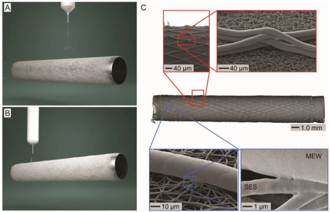
23. Coaxial Bioprinting of Organ Prototypes – From Nutrient Delivery to Vascularization
Introduction:Professor He Yong’s team from Zhejiang University summarized the latest research progress of coaxial bioprinting, detailing the basic principles, technical characteristics, and recent attempts to construct nutrient networks, especially vascularized large-scale structures using this technology, which provides the possibility for rapid manufacturing of vascularized tissue/organ prototypes. The paper “Coaxial 3D bioprinting of organ prototyping, from nutrients delivery to vascularization” was published in the Journal of Zhejiang University-SCIENCE A.— Click ImageView Details —