
Recently, Professor Hu Yong’s team from the Department of Hand Surgery/Ankle Surgery at Shandong University Second Hospital successfully applied 3D printing technology to perform a total talus prosthesis combined with ankle joint replacement surgery for a patient with talus osteonecrosis and ankle arthritis, maximizing the preservation of the patient’s ankle joint function. The patient utilized 3D printing technology comprehensively in the design and production of the prosthesis and trial model, preoperative simulation, and intraoperative osteotomy positioning. According to the literature, there have been no reports on this.
In July 2021, a 36-year-old man, Mr. Wang (pseudonym), accidentally fell and suffered pain, swelling, and bleeding in the right ankle and wrist, with limited movement. He was diagnosed at another hospital with “open comminuted fracture and dislocation of the right talus, fracture of the medial malleolus of the right tibia, fractures of the right cuboid and part of the metatarsal bases, and fracture of the distal end of the right radius.” After surgery, he developed joint infection and experienced pain in the right ankle when walking, severely affecting his daily life. Seeking further treatment, Mr. Wang came to the Department of Hand Surgery/Ankle Surgery at Shandong University Second Hospital. CT and MRI examinations indicated right talus osteonecrosis with ankle arthritis.
Talus osteonecrosis is a common complication of talus fractures. Mr. Wang also had a dislocation of the talus, and conservative treatment was ineffective, necessitating surgical intervention. After thorough discussion, Professor Hu Yong’s team decided to use 3D printing technology to customize a biological total talus prosthesis for Mr. Wang in conjunction with ankle joint replacement surgery.
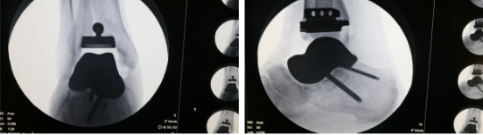
Before the surgery, Professor Hu’s team held multiple discussions, extracted CT data, performed digital three-dimensional reconstruction, designed computer plans, printed trial models and prototypes, designed the prosthesis, and conducted full talus 3D printing to prepare thoroughly for the surgery.
The surgery was performed by Hu Yong and Cao Songhua, with close cooperation from the anesthesia department and the second surgical department. The team thoroughly cleaned the necrotic talus and surrounding inflammatory tissue for Mr. Wang, implanted the trial model of the 3D printed talus prosthesis, and after confirming its suitability, implanted the biological 3D printed prosthesis, securing the talus to the calcaneus with screws. A 3D printed guide was then used for osteotomy at the distal tibia, and after osteotomy, the trial model was inserted, followed by the appropriate tibial ankle joint prosthesis. The surgery was completed successfully. The use of the 3D printed tibial osteotomy guide significantly shortened the surgical time, increased osteotomy precision, and reduced radiation damage caused by fluoroscopy.
Talus osteonecrosis is one of the most common and disabling complications of talus fractures, severely affecting the patient’s walking function and daily life. Traditional treatment methods include ankle arthrodesis or tibio-talar fusion, which, while relieving pain, sacrifice the range of motion of the ankle joint, greatly impacting the patient’s walking function. 3D printed total talus replacement and ankle joint replacement can relieve pain while maximizing the preservation of the patient’s ankle joint mobility, allowing for a better quality of life.
After treatment with 3D printed total talus combined with ankle joint replacement, Mr. Wang achieved better ankle joint function, greatly improving his quality of life. This represents another technological breakthrough for the Department of Hand Surgery/Ankle Surgery following total ankle joint replacement and 3D printed total talus replacement.
Department Introduction
The Department of Hand Surgery/Ankle Surgery at Shandong University Second Hospital is a leading unit in the Foot and Ankle Surgery Group of the Orthopedic Branch of the Shandong Medical Association, as well as the Foot and Ankle Medicine Professional Committee of the Shandong Research Hospital Association. It is the leading unit of the Shandong Foot and Ankle Surgery Specialty Alliance. The ankle surgery team is one of the best in the country, innovating various new surgical techniques in foot and ankle surgery, receiving praise from domestic and international experts; every year, many doctors from within and outside the province come for advanced studies, cultivating numerous specialists in foot and ankle surgery for Shandong Province. We are on a new journey, always moving forward. Standing at a new historical starting point, the Department of Hand Surgery/Ankle Surgery will continue to improve itself, innovate, and contribute to the hospital’s high-quality development.
 Expert Introduction

Hu Yong, Chief Physician, Director of Hand Surgery/Ankle Surgery, Master’s Supervisor. Specializes in the treatment of foot and ankle diseases and trauma. He is at the forefront of treating congenital toe deformities and hallux valgus in China.

Cao Songhua, Associate Chief Physician, Deputy Director of Hand Surgery, specializes in minimally invasive repair and reconstruction of ligament and cartilage injuries in the wrist and ankle joints, as well as arthroscopic minimally invasive treatment of complex fractures in the wrist and ankle.

Authors: Xu Wenpeng, Cao Songhua
Reviewed by: Hu Yong
Source: Hand Surgery/Ankle Surgery
Editor: Lin Yu
Proofread by: Zhang Ruixue, Wang Houjiang
