Training Notification for Multi-SIM AXR Multimodal Super-Resolution Confocal Microscope

Training Notification for Multi-SIM AXR Multimodal Super-Resolution Confocal Microscope

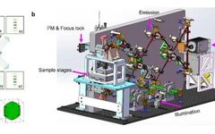
Introduction The Multi-SIM AXR multimodal super-resolution confocal microscope (A23000107) is a microscopic imaging system that integrates structured light illumination super-resolution (Multi-SIM) and confocal (NIKON AXR): Multi-SIM has various super-resolution imaging modes that allow for three-dimensional fast super-resolution imaging, especially suitable for long-term super-resolution imaging of live cells. The Nikon AXR scanning confocal features a 25 … Read more

RNA-Activated CRISPR/Cas12a Nanorobots Operating in Live Cells

RNA-Activated CRISPR/Cas12a Nanorobots Operating in Live Cells

Abstract The active clustered regularly interspaced short palindromic repeats (CRISPR/Cas12a) system possesses cis-cleavage (targeting) and trans-cleavage (off-target) activities, which are highly useful for genome engineering and diagnostic applications. Both single-stranded and double-stranded DNA can activate crRNA−Cas12a ribonucleoprotein (RNP), enabling its cis and trans cleavage enzymatic activities. However, whether RNA can activate the CRISPR/Cas12a system and … Read more

Application of Nanolive in Ferroptosis Research

Cell biologists observe various gene-determined, active, orderly forms of cell death during development, known as programmed cell death, including apoptosis, autophagy, pyroptosis, and ferroptosis. Ferroptosis is a metabolic form of non-apoptotic cell death characterized by iron-dependent lipid peroxidation, and it has garnered significant attention due to its strong correlation with human diseases such as neurodegenerative … Read more

4Pi-SIM Imaging Technology for Live Cell 3D Resolution

4Pi-SIM Imaging Technology for Live Cell 3D Resolution

Subcellular organelles in cells often have intricate three-dimensional structures and biological functions. Observing the three-dimensional distribution and dynamics of organelles using fluorescence microscopy helps understand cellular functions and reveals interactions between organelles. However, fluorescence microscopy is limited by optical diffraction and the numerical aperture (NA) of the objective lens, resulting in blurred low-resolution images. Due … Read more

Using Holographic 3D Imaging to Observe Virus-Induced Cytopathic Effects

Using Holographic 3D Imaging to Observe Virus-Induced Cytopathic Effects

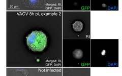
Cytopathic effects (CPEs) are a hallmark of infection. Due to the phototoxicity of classical optical microscopy, CPEs are difficult to observe. This study reports different patterns of viral infection in live cells using Digital HoloTomographic Microscopy (DHTM). DHTM is label-free and records the phase shift of low-energy light passing through the sample on a transparent … Read more