What Exactly Is an ADC?

What Exactly Is an ADC?

Known as magic bullets, ADCs have been highly praised in recent years. The design concept of these drugs is clear: to utilize the high specificity of antigen-antibody binding to selectively deliver toxins to antigen-positive cells, thereby selectively killing target cells and reducing damage to normal cells. However, like other drugs, the actual performance of ADCs on the battlefield may not completely align with the original design concept.

With the emergence of more and more data, the true working mechanisms of successful ADCs are becoming clearer. They represent a highly complex class of drugs, so no single study can fully describe the mechanisms of ADCs. Just like the story of the blind men and the elephant, we need to integrate many research findings to obtain a relatively complete picture.

In this article, I will summarize some important recent data points, allowing readers to connect these points and observe what kind of anticancer drugs are being seen.

1
What Exactly Is an ADC?
ADCs Are Evolving from Molecular Targeting to Tissue Targeting
ADCs are essentially prodrugs with no inherent activity (except for antibody-mediated effects) and only gain cytotoxicity after the intracellular release of toxins. This distribution order is the opposite of traditional small-molecule drugs (which first contact normal cells and then distribute to tumors), providing an inherent advantage for tumor killing. More importantly, this activation strategy allows for targeted killing; if the target antigen is expressed with sufficient specificity, ADCs carrying super-killers can kill tumor cells precisely with very low doses without disturbing the normal cells’ daily life.
However, highly specific tumor antigens are rare, and they are somewhat expressed in normal tissues. Tumors are relatively small compared to the human body, so most antibodies at low doses are primarily absorbed by normal tissue targets. This is also why diagnostic antibody imaging agents usually need to be used in conjunction with antibodies, and some people use monoclonal antibodies to solve binding site barriers.
Additionally, non-specific binding of antibodies and endocytic forces are rampant; after all, antibodies cannot wander indefinitely in the system, so the body has evolved a whole set of antibody metabolism and clearance mechanisms that do not rely on antigens. ADCs are not exempt from this and will also be degraded and cleared at specified times and locations. Another very important factor is that although tumor antigen expression may be high, the antigens available for drug interaction are usually limited due to the disrupted vascular structures and tight junctions between cells in the tumor tissue. Using antibody imaging agents to diagnose tumors has high practical value, but so far, apart from a few imaging agents, antibody-based imaging agents have not been marketed for use.
One reason is that tumor antigens have limited specificity, leading to significant background noise. Another important reason is that tumor tissue is too chaotic, making it difficult for large molecules to penetrate. Trastuzumab imaging agents fail to visualize in 1/3 of HER2-positive lesions, while accumulating significantly in non-HER2 tissues such as the liver, kidneys, and spleen. One can imagine the treatment window if the payload carried by trastuzumab were not an imaging agent.
The concealment of tumor antigens, combined with the interference from the binding of antibodies to normal tissues, results in insufficient navigation capabilities for current ADCs, leading to over 99% of ADCs being unable to locate tumor tissues, forcing them to release toxins in normal tissues. A fundamental difference between ADCs and small-molecule prodrugs is that ADCs cannot be excreted by the kidneys due to their large molecular weight and must be degraded to release toxins.
An ADC with a half-life of one week means that within that week, regardless of how many antigens it finds, it must release 50% of its toxins, much like a city’s waste station with limited capacity that is hard to find, where citizens cannot avoid the garbage, making it difficult to ensure the city’s appearance. Even in the microenvironment, tumor cells are not always the only cells releasing toxins; many non-specific release mechanisms may participate in the toxin release of ADCs, which also diminishes the therapeutic value of target-mediated effects.
Therefore, highly toxic ADCs do not succeed; it is only after the appearance of less toxic toxins like DXd that ADCs truly entered the mainstream. For membrane-permeable toxins, intracellular release does not equate to intracellular residence but instead spreads to other cells in the microenvironment. If released in tumor tissues, this would extend the lethality to tumor cells that do not express target antigens, known as bystander killing, thus enhancing efficacy.
However, this property is a double-edged sword; toxins released in normal cells will also spread to other normal cells, causing a larger range of damage. One cannot expect to socialize tumor killing while capitalizing on normal tissue damage. Clinically, ADCs have rare target and off-target toxicity; the same toxin, regardless of the ADC it is made into targeting any antigen, has similar toxicity profiles and maximum tolerated doses, indicating that regardless of where the toxin is released, the final toxicity is similar and will be socialized across the entire compartment. DXd ADCs cause hair loss, while MMAF ADCs cause ocular toxicity. Of course, it is possible that ADCs with target toxicity were eliminated during development, but based on my understanding of preclinical evaluation systems, this possibility is not very high.
The situation regarding efficacy is similar; although some ADCs benefit more in populations with high antigen expression, most ADCs show weak correlation between response and target expression in most application scenarios. Most ADCs lack reliable predictive biomarkers for efficacy; currently, 15 ADC products have been launched globally, with only three requiring target antigen testing to select patients. Some high-density antigens like HER2 may also be expressed in negative tumors, so there are opportunities for entry into cells, but normal tissues such as the kidneys can express HER2 at IHC 3+ levels. This is not targeted delivery but rather treating normal tissues as human shields (of course, different toxins have different killing abilities against different normal tissues); if magic bullets work on both normal tissues and tumors, it would be better to directly deliver DXd intravenously.
This outcome may stem from several theoretical possibilities. First, the diffusion speed of small-molecule chemotherapy drugs is usually much faster than their cell-killing speed. Traditional chemotherapy drugs are generally infused into a patient’s vein, but the drugs do not stay in the blood for long; instead, they distribute to body tissues in a short time, meaning that intravenous chemotherapy cannot be considered targeted blood drugs. Similarly, the toxins released by ADCs typically diffuse into the microenvironment before killing the tumor cells that release them, which is one reason why intratumoral injection of chemotherapy does not better control tumors.
If the cells releasing the toxins must be killed before the toxins can continue to kill other cells, then antigen-positive cells should be prioritized for elimination, and the remaining tumor cells should predominantly be target-negative. However, in the DAISY clinical trial of Enhertu, patients who responded showed only a slight decrease in HER2 expression after progression, and many patients had no change in HER2 levels, with some even showing an increase in HER2 levels; this behavior of acting against the wind indicates that DXd performed indiscriminate killing in the microenvironment.
Second, the binding of ADCs to antigens is only one step in achieving targeted delivery; subsequent steps must also cooperate to realize the value of this delivery step. For example, endocytosis must be fast enough to enter lysosomes, linkers must be cleaved, and toxins must escape from lysosomes; otherwise, cells have a series of operational mechanisms that can expel ADCs, such as FcRn-mediated antibody recycling. Therefore, the binding of ADCs to antigens is a relatively fragile union, and many trivial matters can cause this uniquely selective delivery event to fail.
In summary, although ADCs are designed as molecular targeting delivery technologies, under the guidance of clinical treatment windows, they have evolved into tissue-level delivery technologies, where both efficacy and toxicity are at the tissue level, determined by the tissue’s free toxin AUC or Cmax. Of course, membrane-impermeable toxins or non-cleavable ADCs are different, but such ADCs are increasingly rare. This macro picture has profound implications for ADC target selection and molecular design.
2
What Exactly Is an ADC?
ADCs Are Not Magic Bullets, But Magic Triggers
ADCs are referred to as magic bullets, meaning that chemotherapy drugs linked with antibodies can accurately kill target cells.However, this is based on the assumption that antibodies can specifically bind to antigens, allowing toxins to specifically kill antigen-positive cells.In reality, the binding of antibodies to antigens is just a small step; if endocytosis is slow, ADC drugs may still dissociate from the antigens. Even if endocytosis is efficient, if the linker hydrolysis is too slow, ADCs may still be expelled from the cell by FcRn.
Relative to normal tissues, tumors are just a tiny area, so ADCs spend more time interacting with normal cells. Normal cells also have mechanisms for endocytosing ADCs and releasing toxins, such as Fc receptors. Although individual normal cells may be less efficient than tumor cells due to the lack of target antigen expression, normal cells dominate in number. Coupled with the more intact vascular structure of normal tissues compared to tumor tissues, drug penetration is better, meaning that less than 1% of ADCs are released in tumors.
Even if ADCs can release toxins in tumor cells, achieving selective killing still requires that the toxins remain in the target cells long enough. Small molecules have strong diffusion capabilities, and tumor cells also express some efflux pumps, so for membrane-permeable toxins like DXd, no matter where they are released, they must gather in the playground and then act collectively to kill tumor cells in the microenvironment. The working mechanisms of less membrane-permeable toxins like DM1 may differ.
If we compare ADCs to paratroopers, non-membrane-permeable T-DM1 completes its killing task upon landing in the target cell, while T-DXd lands in the enemy’s backyard and then acts collectively, allowing it to simultaneously kill both antigen-positive and negative tumor cells until expelled from the body. As mentioned earlier, this mechanism in the tumor microenvironment results in bystander killing effects, while in normal tissues, it leads to off-target toxicity.
Thus, what ADCs can truly achieve is a certain degree of selective toxin release in antigen-positive cells; their working mechanism resembles a magic trigger, meaning that under certain circumstances, they can pull the trigger in tumor cells, but whether they can become magic bullets depends on how long the bullets fly and where they land.
Two years ago, scientists from Zymeworks published a significant article pointing out that ADCs do not show improved absolute safety compared to small-molecule toxins; the maximum tolerated dose (MTD) has not increased. Logically, if ADCs only fire at terrorists, bystanders should not worry about being hurt, but the actual situation is that the proportion of bystanders hurt remains unchanged compared to small-molecule toxins. Not only has the number of bystanders not decreased, but those hurt are still the same unfortunate group, such as rapidly dividing blood cells, which are victims whether facing ADCs or small-molecule toxins.
This core viewpoint was proposed as early as 2015 by FDA scientists, stating that the toxicity strength and spectrum of ADCs are fundamentally determined by the toxins, regardless of the target. The reason is that although the bullets are primarily fired in tumor cells, how long they fly and where they ultimately cause damage is determined by the AUC of the toxins in each compartment.
Although absolute safety has not improved, it does not mean that ADCs do not provide additional benefits. When the blood drug concentration of toxins reaches MTD, the concentration of toxins in the tumor microenvironment may vary depending on the level of antigen expression, thus possibly improving absolute efficacy. It is challenging to study how much ADCs accumulate in tumors compared to small-molecule toxins, especially in the actual tumors of patients.
Efficacy cannot serve as a substitute indicator for toxin accumulation; first, different tumors have different sensitivities to the same toxins, and tumors with high antigen expression may be more sensitive or more tolerant to toxins. Second, it is challenging to eliminate the influence of the antibody component; for tumors with high target expression, antibodies themselves may mediate some immune killing, making the additive effects challenging to quantify. This ties in with the third factor: the slow-release mechanism, as ADCs are a slow-release toxin mechanism, which also increases the therapeutic window.
One method is to attach various imaging agents to antibodies to see where the imaging agents remain, thereby inferring the possible release mechanisms of toxins, but these technologies are challenging to precisely target cell types. Some studies have indeed pinpointed cell types, but the results can be awkward. For example, a trastuzumab imaging agent primarily binds to macrophages in HER2-positive solid tumors, not targeting HER2; there are various possibilities for why highly specific trastuzumab has become a poor match.
Of course, one can directly measure drug concentrations in tumor tissues, which also has several confounding factors. First, if measuring total toxin concentration, one must consider the drug-protein binding rates in different tissues; for instance, some small-molecule toxins like MMAE may accumulate in tumor tissues compared to blood but do so through non-specific binding to proteins, meaning free drugs have not accumulated. Additionally, small molecules can easily escape from tumors, so even if many toxins are released in tumors, rapid escape can lead to decreased AUC, which interferes with fast-acting or slow-dissociating (slow Koff) toxins.
This year, scientists from AstraZeneca published data on drug release from Enhertu in mouse models, showing that the AUC of DXd released by Enhertu in HER2 high-expressing tumors (N87, with each cell expressing 3.5 million HER2) was 493, while in HER2 low-expressing MB468 (with each cell expressing 4,800 HER2 receptors), it was 156. From a navigation capability perspective, HER2 should be seen as the ceiling for ADC targets; the threefold difference in toxin accumulation between strongly positive tumors and nearly negative tumors reflects the limits of this delivery mechanism under the current ADC design framework.
While the threefold enrichment did not meet many people’s expectations, overcoming thermodynamic distribution laws is not an easy task. The therapeutic window for chemotherapy is usually very small, and a threefold increase is still a significant improvement. Coupled with the auxiliary role of the antibody component, especially in populations with high target expression and the slow-release mechanism of ADC toxins, Enhertu significantly improved therapeutic effects compared to both trastuzumab and irinotecan.
However, ADCs are also a class of drugs that are highly complex in design, production, and development. The question remains whether this threefold enrichment justifies such a complicated design and whether it can be achieved through simpler designs. If one is willing to bear the high costs associated with antibody conjugation, we need to rethink how to truly achieve a significant improvement over simpler molecular construction models. ADCs have reached a point described by Deming: survival isn’t mandatory; they either reinvent themselves or be replaced by other models. Both choices present new opportunities.
3
What Exactly Is an ADC?
IO-Chemotherapy, Even IO-IO Combinations
For the designers of magic bullets, a more humiliating statement than magic triggers is that your weapon is merely a combination of gunpowder and a gun handle, and the aiming and navigation systems are not as important as you think. However, truly successful ADCs have indeed relied on the synergistic or additive effects of both the antibody and toxin components.This combination effect has been relied upon in tumor treatment throughout history and has a complex background.
The greatest obstacle in tumor drug development is the issue of tumor heterogeneity. If every tumor cell had the same structure and function and the same relationship with drugs, curing advanced tumors would have long been a reality. Tumors exhibit both genetic heterogeneity, meaning that each tumor cell’s genome varies and expresses key proteins in different structures and quantities, and spatiotemporal heterogeneity, meaning that protein expression varies among tumor cells at different developmental stages and regions. The infiltrative regions of drugs also differ among lesions, and the existence of tumor stem cells makes treating advanced tumors almost an impossible task.
Because of tumor heterogeneity, most therapies can only kill part of the tumor; targeted therapies struggle to kill target-negative tumors, and immunotherapies lack the support of immune police in the microenvironment. Small molecules have poor selectivity, and large molecules cannot effectively infiltrate the microenvironment. Even chemotherapy struggles to kill tumor stem cells and cannot effectively prevent tumor regeneration.
Therefore, combination therapy is the universal strategy for almost all tumor treatments. Even relatively clean targeted therapies can only effectively control advanced tumors in a few special scenarios when combined with chemotherapy; currently, most treatment regimens involve chemotherapy. Most targeted therapies must be used in conjunction with chemotherapy to truly demonstrate therapeutic value, and any new treatment regimen still needs to bear the shadow of chemotherapy. Chemotherapy drugs can kill a batch of stubborn molecules, but the cost is that they may harm some innocent bystanders. They can also create enough chaos in tumor cells and the tumor microenvironment to drive gangsters out of their hideouts, facilitating more precise and gentle modern therapeutic targeting, thus providing a basis for efficacy.
The discovery and development of chemotherapy drugs primarily focus on their killing ability, but in real patients, they do not necessarily achieve this through direct killing. Most chemotherapy drugs that show sufficient therapeutic windows in clinical trials do not directly kill tumor cells at tolerated doses because tumors in patients grow much slower than those in laboratories. One hypothesis is that chemotherapy drugs cause significant chaos within tumor cells before reaching lethal doses, thereby activating the immune system to indirectly kill tumors.
This is akin to a hitman entering a gangster’s gathering place to clean up the gang; although he possesses combat capabilities, he can create chaos in the tavern to attract police to surround the previously undetected gangsters. There have long been rumors that chemotherapy contains elements of immunotherapy, and now IO-ADC combinations are also an important direction in clinical development.
Antibody drugs must also leverage the immune system. Most antibody drug targets are expressed at higher levels in tumor cells but do not necessarily participate in crucial decisions for tumor development. Of course, higher expression than normal cells is also a suspicious sign, possibly indicating more covert involvement. Even targets like HER2, which are highly related to tumor growth, have limited effects on tumors by blocking signaling pathways; the impact is more through immune-mediated effects like ADCC and ADCP. Therefore, ADCs in the tumor microenvironment have both antibody-mediated immune killing and direct killing from chemotherapy or indirectly mediated immune killing, simulating a combination of chemotherapy and antibody drugs in certain scenarios.
Of course, some ADCs have low MTDs, and the antibody component does not reach therapeutic doses, but successful ADCs achieve effective antibody doses. Some ADCs do not have significant single-agent activity from the antibody component, but the local drug combination in the tumor microenvironment may still produce therapeutic effects. Research from decades ago has shown that a drug can sensitize another drug’s therapeutic effect at far lower than therapeutic doses, which is an important theoretical basis for designing small-molecule dual-target drugs; thus, the contribution of the ADC antibody component should not be easily overlooked.
As mentioned earlier, most ADCs show weak correlation between response and antigen expression in most scenarios, and ADCs made with the same toxin show very similar toxicity and toxicity profiles regardless of target distribution, indicating that target-mediated biological effects primarily manifest at the tissue level rather than at the cellular level. Some ADCs show a certain target-related response and efficacy, such as Enhertu, where HER2 high-expressing patients respond better. However, it is difficult to distinguish whether this is due to higher tumor toxin release throughput or greater sensitivity to trastuzumab-mediated immune killing.
In animal models, toxin accumulation in HER2 high-positive tumors is several times higher than in nearly non-expressing tumors, but patient tumors may be more complex, leading to different results. HER2 imaging agents can distinguish between positive and negative lesions to some extent, but the false-positive and false-negative rates are still high and do not reach commercial application levels. Additionally, as mentioned earlier, the accumulation of antibodies in antigen-positive cells does not equate to the release of toxins in those cells, and even if released, the toxins may not remain long enough.
Enhertu has been approved for the treatment of all HER2-positive solid tumors, but the response rate of 70% in HER2-positive breast cancer starkly contrasts with the 5% response rate in HER2-positive pancreatic cancer. This discrepancy seems more related to the basic efficacy of trastuzumab in these two types of tumors rather than the navigation capability of Enhertu, as both are HER2-positive. Of course, the sensitivity of these two tumors to DXd and their toxin release abilities may differ.
Tumor treatment has always relied on combination therapies anchored by chemotherapy. ADCs are the only single-agent drugs that possess both the broad-spectrum killing of chemotherapy and the targeted killing of antibodies. Therefore, ADCs are the only ones capable of launching a three-dimensional war against tumors, aligning with years of experience in combination therapies against cancer. Unfortunately, many antibody targets for ADCs do not have marketed monoclonal drugs; one reason may be that these antibodies also require suitable chemotherapy combinations to truly take effect, while marketed chemotherapy drugs either do not match the mechanism or have non-parallel pharmacokinetics, leading to low combination hit rates.
In fact, there are very few antibody drug targets with significant single-agent activity in solid tumors. I counted and found that only EGFR, HER2, PDL1, CLDN18.2, VEGFR2, PDGRFα, and EpCAM have marketed drugs, most of which are small products. Therefore, any antibody with some single-agent activity should be made into an ADC to test its druggability. The popularity of ADCs has resulted in clinical verification of which toxins are quality partners for antibody combinations, such as DXd, which seems to perform well with multiple antibodies, at least under slow-release conditions. These valuable data provide technical support for the rapid validation of new target ADCs.
4
What Exactly Is an ADC?
Curtin-Hammett Rule
A fundamental assumption in ADC design is that the binding of antibodies to target antigens will initiate the killing process; if ADCs bind only to targets, they will only kill cells expressing those targets, and if only tumors express the targets, ADCs will only kill tumor cells.Of course, in practice, this logical chain often breaks down, as the human body does not have antigens expressed solely on tumor cells, and tumor surface antigens are not all accessible to ADC drugs.Normal cells that do not express antigens can also endocytose large amounts of ADCs and release toxins; even within the tumor microenvironment, how much ADC toxin is released through antigen-positive tumor cells remains uncertain.
These little dirty secrets will be discussed slowly in the future. Today, let’s talk about how ADCs must achieve tumor-specific killing with the right timing, location, and conditions.
Chemical reaction kinetics provide some theoretical foundations for predicting the final outcomes of multi-step processes, and one such rule, the Curtin-Hammett rule, is particularly relevant for understanding the final outcome of ADCs killing tumor cells in this multi-step process. This rule states that the product ratios of a chemical reaction with different pathways are determined by the activation energies of each pathway, independent of the concentrations of intermediates, provided that the interconversion speeds between intermediates are fast.
Let me translate this into a life scenario to help non-chemistry professionals understand. Imagine two restaurants, A and B, on a street, each employing a person to attract customers outside. The person at restaurant A is more eloquent, so 90% of the foot traffic is drawn to their restaurant, but does this guarantee that restaurant A’s revenue is higher than restaurant B’s? Not necessarily; if restaurant A has a dirty, chaotic dining environment, poor service, and bad food, most customers may end up dining at restaurant B, which is just next door. Of course, if restaurant A is in Beijing and restaurant B is in Shanghai, this rule fails, corresponding to a chemical reaction where the interconversion speeds between intermediates are slow.
The same principle applies to the binding of ADCs to target antigens. Typically, the speed at which antibodies bind to antigens is quite fast, often reflecting the diffusion speed of the molecules, but whether they can be endocytosed into the cells depends on the biological functions of each antigen and the construction mode of the antibodies. Once inside the cell, they do not immediately unload their toxins; instead, they must reach a specialized department rich in degrading enzymes called lysosomes to unload, and the quantity and activity of tissue proteases within lysosomes differ in different tumor cells, leading to varying hydrolysis speeds of linkers.
The time spent in lysosomes is limited; if the hydrolysis speed is too slow, ADC molecules may be expelled from the cell by mechanisms such as FcRn recycling, requiring them to re-enter the queue to return to lysosomes. The proteases within lysosomes do not only degrade linkers; if the toxins resemble peptides, they may also be treated as waste. Previously, Velcade was used as an ADC toxin, but it was treated as a peptide and broken down within lysosomes.
Even if your ADC successfully releases all its toxins in the target cells without having to queue repeatedly, this does not mean the assassination task is complete, and they can claim their reward; because relative to escaping tumor cells, tumor killing is a slow process. Once toxins are released, they lose all connection to antibodies and diffuse randomly, going off on their own. Some toxins, due to their slow membrane permeability, may remain in tumor cells for an extended period, providing enough time to kill that tumor cell, but the cost is that they can only kill that specific tumor cell as they cannot enter other cells.
Currently, most ADC toxins can produce what is known as bystander killing effects, meaning they can freely enter and exit cell membranes, with the timing of this entry and exit measured in seconds. However, tumor cell killing also takes days in vitro, and patient tumors grow much slower, so even the most sensitive tumors may take weeks to show a response.
Thus, the timing of toxin release from tumor cells and the killing of tumors do not align. Except for highly active ligands or irreversible ligands (with a very high dissociation constant, Koff, requiring a long time to dissociate from the target), most small-molecule toxins, even when released at their destination, scatter before completing their tasks.
Like all targeted delivery technology platforms, ADCs hope to maintain drug concentrations above systemic exposure levels in the tumor microenvironment for extended periods, but this violates the second law of thermodynamics. To stay away from thermodynamic equilibrium, continuous energy input is required, and this energy input comes from the binding energy between antibodies and antigens. However, this is merely a necessary but insufficient condition; as analyzed above, the efficiency of this limited binding energy usage is related to many factors, and if misused, it can easily lead to financial strain. Similarly, the binding of targets to ADCs is also a necessary but insufficient condition; increasing marriage rates does not equate to increasing birth rates, as the latter is also dependent on many other factors.
5
What Exactly Is an ADC?
Magic Bullets or Just Bullets
Although ADCs are much larger and more complex than chemotherapy drugs, they are essentially still chemotherapy drugs.While the antibody part acting as a driver may play a covert protective role in certain cases, the killing still primarily depends on the toxin component; magic bullets are still just bullets.Despite the long-standing concept of ADCs and the rapid emergence of heavyweight products like T-DM1, ADCs did not receive significant attention from mainstream pharmaceutical companies until the advent of Enhertu.
In 2020, Roche’s CEO stated, “We have shifted our technology priorities,” Roche CEO Severin Schwan told Reuters. “Maybe others will be luckier, but we failed to master the complexity.” Roche’s position in the oncology drug field is akin to Apple’s in the smartphone sector; at that time, T-DM1 and Polivy were already marketed, and what Roche’s leader said would certainly be taken seriously by the industry. In retrospect, it can be said that everything was ready except for a quality toxin, and that quality toxin was DXd.
Taiho Pharmaceutical had been developing camptothecin derivatives for many years and launched irinotecan. While irinotecan has good efficacy, it still falls short compared to taxanes and platinum drugs. Taiho then discovered a more active analogue, exatecan, but with greater toxicity; despite conducting numerous clinical trials, they could not find a usable indication. At that time, Taiho aimed to create a polymer conjugated prodrug of exatecan and advance a polymer prodrug called DE310 into clinical trials.
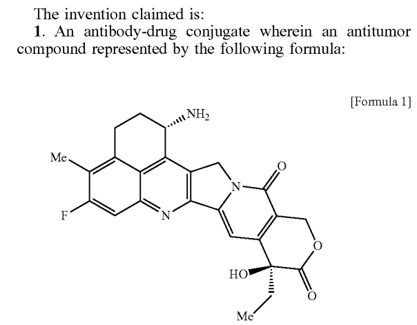
This prodrug released exatecan very slowly, but it still could not achieve a sufficient therapeutic window. Later, as ADCs entered the spotlight, Taiho transferred the experience from DE310 to ADC design, including the GGFG linker used in DE310. Even the earliest patent for Enhertu was based on exatecan as the toxin:
What Exactly Is an ADC?
While exatecan is more active than SN38, it is still considered relatively weak as an ADC toxin; this drawback can be mitigated by a high DAR value. Exatecan has an amino group that can directly couple with GGFG through an amide bond, but this coupling can lead to some breaking at the G-F junction during enzyme cleavage, releasing G-exatecan. Considering some physicochemical properties, Taiho’s scientists added a hydroxyethylamide group to exatecan, which is now famously known as DXd (deruxtecan).
Before this, how GGFG coupled with hydroxy compounds was already known technology, so making Enhertu was not complicated. DXd is approximately five times weaker than exatecan, which does not align with the mainstream ADC design philosophy at the time; however, no one is currently concerned about the in vitro activity of DXd.
The success of Enhertu was somewhat unexpected, as evidenced by Taiho’s stock performance over the past 20 years. As for why Enhertu has been so successful, especially in HER2 ultralow breast cancer, there is still research ongoing in the industry. However, it is estimated that DXd possesses certain unique superpowers. Currently, at least ten other HER2 ADC products using different toxins have failed in clinical trials, as have other more lethal candidates such as CAR-T, CAR-M, and immunotoxins. Meanwhile, DXd’s five different target ADCs are each worth a fortune, having been transferred to AstraZeneca and Merck for a total transaction value of $35 billion.
As for the mysterious capabilities of DXd, it may take a long time to clarify; it could simply be that it has a higher safety window than exatecan and is itself a quality small-molecule chemotherapy drug. Unfortunately, DXd has never entered clinical studies, so there are no head-to-head comparison data. It is also possible that it produces some immune activation effects at non-killing doses or directly induces ICD (immunogenic cell death) or sensitizes antibody-mediated ADCC and other immune effects.
DXd’s short half-life is a disadvantage for small-molecule drugs but an advantage for ADC toxins, as toxins entering the bloodstream and passing through metabolic and excretory organs such as the liver and kidneys will be quickly eliminated, maintaining a drug gradient between tumor tissues and the bloodstream. Perhaps it is this combination of factors that makes it challenging to assess with the current limited data.
The discovery of new drugs is now a super complex system aimed at reducing clinical failure risks through a series of so-called de-risking screening processes, avoiding the trial-and-error approach of clinical trials. Unfortunately, true innovative new drugs are still primarily identified through clinical trials, making rational drug design a field that relies heavily on chance. Even so-called me-too follow-up drugs may be easier, but it depends on specific situations; for example, important drugs like taxanes, proton pump inhibitors, thalidomide-type molecular gels, and P2Y12 receptor agonists have only very slight structural differences from their original drugs.
This indicates that mechanisms alone are insufficient to filter for quality drugs; otherwise, someone would have already screened for new structural types. Many mainstream anticancer drugs used for decades still leave us unclear about their true working mechanisms, such as paclitaxel, which, at clinically effective doses, does not sufficiently inhibit microtubule protein disassembly. Other important drugs like platinum agents and tamoxifen face similar issues. DXd is merely the latest enigma, and the more such enigmas, the better.
6
What Exactly Is an ADC?
Who Is Casting the Spell?
ADCs themselves have no inherent activity (except for antibody-mediated immune killing); only when the toxins are released do they exhibit tumor-killing activity. However, the details of toxin release in vivo, especially in patients, remain quite murky.Although the design of ADCs aims to release toxins through endocytosis in antigen-positive tumor cells, proteolytic enzymes exist not only in tumor cells but may not even be the primary source.ADCs linkers are primarily hydrolyzed by tissue proteases but may also become substrates for other proteases.Thus, who is casting the spell behind the scenes is not a clear-cut issue.
Although antibody drugs have a 30-year history, there are still many gaps in our understanding of the degradation processes of antibodies in the human body. Studies using isotopic labeling have found that the organs with the highest metabolic degradation efficiency for antibodies in mice are the liver, spleen, and kidneys; however, due to the greater amounts of skin and muscle tissue, the organs that degrade the most antibody protein in absolute amounts are the liver, skin, and muscle, clearing about 75% of antibodies. Many advanced cancer patients experience severe weight loss, and one hypothesis suggests that the dysfunctional degradation mechanisms in muscle may be linked to increased antibody degradation.
It is generally believed that the endothelial phagocytic system is the primary mechanism for antibody degradation, with monocytes and macrophages being the main cell types. This system’s natural biological function is to phagocytize and degrade discarded proteins and other biological materials, giving it a large throughput and strong endocytosis and degradation capabilities, which are also required for ADC toxin release.
There are also large numbers of tumor-associated macrophages (TAM) within the tumor microenvironment, which have typically been co-opted by tumors to aid their growth. TAMs are the most abundant immune cells in the microenvironment and can sometimes outnumber tumor cells. Unlike tumor cells, which are tightly linked in a structured organization, TAMs can move freely within the microenvironment; before becoming accomplices, these cells need to patrol their tissues to clear away waste proteins.
Macrophages seek out culprits based on chemical signals, which is why some have used macrophages as carriers for chemotherapy drugs, leveraging their ability to recognize tumors and other abnormal tissues to deliver drugs to diseased tissues. Drugs can be loaded into macrophages in vitro or stick to macrophages in vivo due to their natural tendencies, a process known as in vivo hitchhiking. You may be familiar with the concept of using red blood cells to deliver drugs, but macrophage delivery is just less high-profile.
Macrophages express various Fc receptors that can bind to the Fc region of IgG antibodies and endocytose the entire antibody. One of the main killing mechanisms of antibodies, antibody-mediated cellular phagocytosis (ADCP), utilizes the ability of Fc receptors to bind and endocytose antibodies; if the variable region binds to tumor cells, the entire complex is phagocytosed. Although Fc receptor-mediated endocytosis is related to Fc conformation and density (which changes upon binding to tumor cell surface antigens), individual antibodies can also be endocytosed by macrophages through Fc receptors.
Although Fc receptors do not bind as strongly to Fc as antibodies do to antigens, the sheer number of TAMs in the microenvironment and their constant wandering means they can significantly impact antibody drugs. PD-1 antibodies, after briefly binding to PD-1, tend to move to the surface of macrophages, which is one mechanism for developing resistance. PD-1 is also a receptor expressed on immune cells, whereas the tumor surface receptors targeted by ADCs may be more significantly influenced by macrophages due to their lower exposure.
Since TAMs can influence the metabolism of monoclonal antibodies, ADCs may also be degraded by TAMs. Seattle Genetics discovered as early as 2017 that some ADC drugs, while targeting tumor antigens, primarily release toxins through macrophage-mediated endocytosis in the microenvironment, with target-mediated release lagging behind the pace of macrophages. Subsequent studies found that if host macrophages were cleared, ADC activity significantly decreased; if monoclonal antibodies blocked ADC binding to tumor cells, ADC levels in the microenvironment decreased, but toxin release remained unaffected. However, if the Fc of silent antibodies was silenced, toxin release slowed significantly.
The significant influence of TAMs on ADC degradation is not only related to their quantity and endocytic capacity but also to the high levels of hydrolytic enzymes in TAMs due to their work requirements. Some studies have used ABPP chemical labeling to trace the sources of active tissue proteases in the microenvironment, discovering that at least in some tumor microenvironments, over 90% of tissue proteases come from macrophages rather than tumor cells. Of course, these observations were made under specific experimental conditions, but they indicate that ADCs in the microenvironment do not only interact with tumor cells.
Animal models are fundamentally different from human tumors; unfortunately, there is very little data on the metabolism and distribution of ADCs in human tumors. Half of this knowledge may come from the DAISY Phase II clinical trial of Enhertu. This trial not only discovered for the first time that Enhertu has a significant response rate in patients with extremely low HER2 expression (recently validated in the DB06 trial) but also conducted very detailed studies on efficacy and resistance mechanisms. Among the studies on PK/PD relationships, it was found that HER2 expression does not significantly correlate with Enhertu accumulation in tumors, and low levels of Enhertu in tumor microenvironments do not rule out the possibility of response.
The authors did not measure the levels of the toxin DXd, nor did they measure the levels of free DXd, so there are no strict PK/PD correlation data. In resistant patients, HER2 levels typically show only minor decreases or no change, with some patients even showing upregulated HER2 levels after developing resistance. These data raise suspicions that there are mysterious forces beyond HER2 involved in the release of Enhertu’s toxins.
Antibodies are highly evolved, multifunctional biological molecules, and toxins have been screened through years of preclinical and clinical trials, eliminating most “poor quality toxins.” ADCs are a highly complex class of drugs, and our understanding of these drugs is still deepening. This series of short articles attempts to depict the image of ADCs from different angles like blind men touching an elephant; what kind of anticancer drugs do you see by linking all these data points?
[Reprint Statement] This article is reproduced from the “US-China Drug Source” WeChat public account.

What Exactly Is an ADC?

Scan the WeChat QR code to add the editor of the Antibody Circle,and qualified individuals can join the Antibody Circle WeChat group!
Please specify: Name + Research Direction!
What Exactly Is an ADC?
The articles reproduced by this public account are for the purpose of conveying more information and clearly indicate the source and author. If any media or individual does not wish to be reproduced, please contact us ([email protected]), and we will immediately delete it. All articles represent the author’s views and do not represent the position of this site.
What Exactly Is an ADC?

Leave a Comment