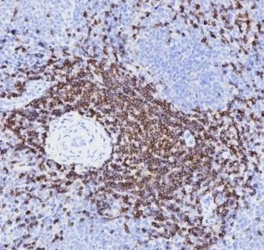
Immunohistochemistry、Immunocytochemistry and Immunofluorescence are three commonly used applications. Do you often feel confused about these three applications? They indeed have many similarities. Let’s clarify their differences.

IF (Immunofluorescence) / IHC (Immunohistochemistry) / ICC (Immunocytochemistry) are three applications, and one part that everyone will definitely not confuse is “Immuno”, because all three applications involve the use of antibodies. First, let’s start with the naming.
Immuno refers to immunological techniques (e.g., the binding of antibodies and antigens).
Histo refers to tissues (cells and their surrounding extracellular matrix).
Cyto refers to cells (excluding the extracellular matrix).
Chemistry here refers to chemical detection methods (e.g., color changes).
Fluorescence refers to the detection of excited fluorophores.
By explaining the roots of these terms, I believe you are no longer confused.
These three application techniques all belong to immunological techniques, visualizing the binding of antigens to antibodies, meaning that through certain methods, the experimental results can be directly observed. The commonly used methods are chemical color development and fluorescence color development. If the report label is enzyme-based, it is immunochemistry; if it is fluorescent, it is immunofluorescence.
In fact, one reason we are confused about immunofluorescence is its English name, Immunofluorescence (IF). However, if we write it as Immunohistofluorescence (IHF) or Immunocytofluorescence (ICF), wouldn’t it be clearer? Although these two terms are not widely used in English literature yet, they are definitely not something I made up; some scholars have already started using them, and standardization is just a matter of time.
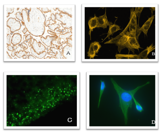
Additionally, their distinctions lie in:
1. Chemical staining methods and fluorescence excitation staining: If it is enzyme-based, it is immunochemistry (IHC/ICC); if it is fluorescent, it is immunofluorescence (IF).
2. Specimen preparation: Immunofluorescence generally uses frozen sections to reduce interference from impurities; while enzyme immunohistochemistry can use either paraffin or frozen sections.
3. Experimental steps: Immunofluorescence staining steps are simpler, while enzyme immunohistochemistry methods are more complex, adding the DAB color development process.
4. Specimen preservation after staining: Specimens stained with immunofluorescence are generally photographed shortly after staining, as fluorescence diminishes over time; while specimens stained with enzyme immunohistochemistry can be preserved for a long time.
5. The results of immunohistochemistry and immunofluorescence can not only indicate whether the protein is expressed more in the cytoplasm or cell membrane but can also be used for semi-quantitative analysis.
6. The images obtained from immunofluorescence are colorful and more visually appealing, suitable for high-quality publications.
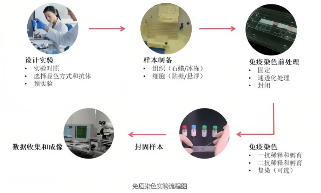
To achieve reliable and consistent experimental results, optimizing the operational process is essential. The principles and experimental steps of IHC/ICC/IF are generally similar, as shown in the overall experimental flow diagram below.
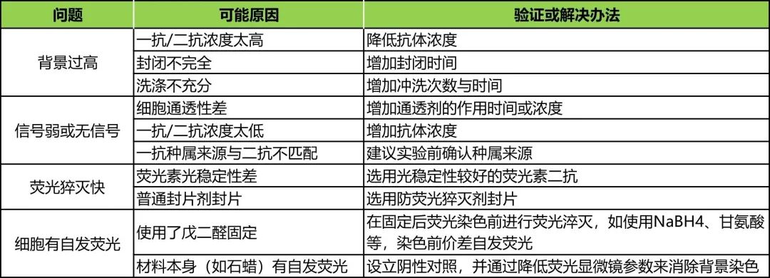
Although the principles of IHC, ICC, and IF are simple, they are often stumbling blocks for many researchers on their long journey to graduation. Here, I present a comprehensive troubleshooting guide to help everyone solve the problems encountered in experiments.
IHC & ICC
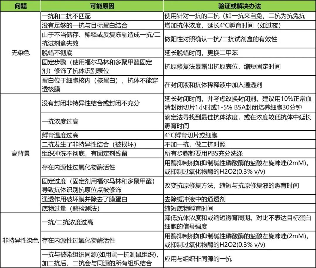
IF
This concludes the current content.
Has everyone mastered it?
See you next time!
[END]

Produced by: Graduate School New Media Center
Edited by: Liu Xiaofei, Liu Xuanyi, Zuo Jinshi, Zhang Ziyan
Proofread by: Yin Bowen, Zhao Siheng