
The Brodmann brain cortex areas were proposed in 1909 by German physician Korbinian Brodmann and have been continuously revised to become the most widely used method of brain partitioning today. Today, we share several beautiful images of the Brodmann brain cortex areas.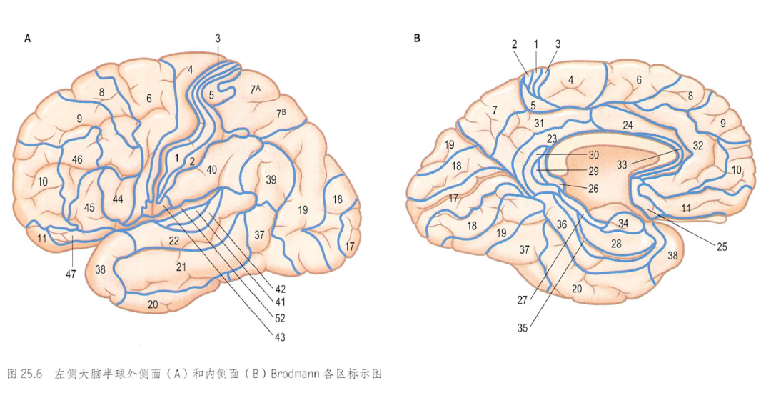
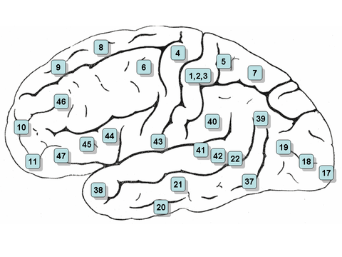
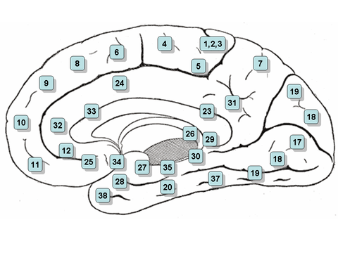
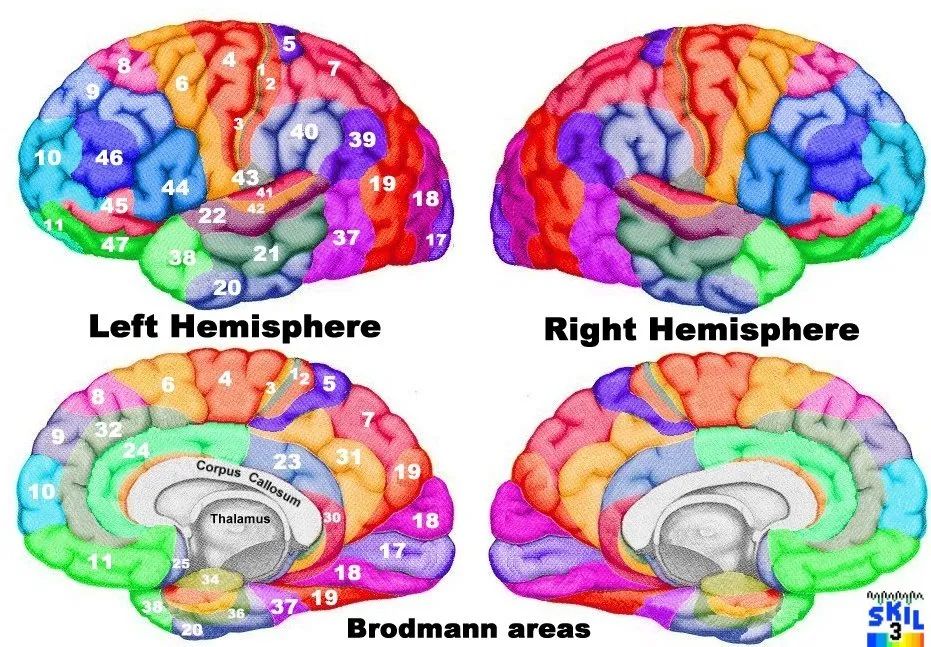
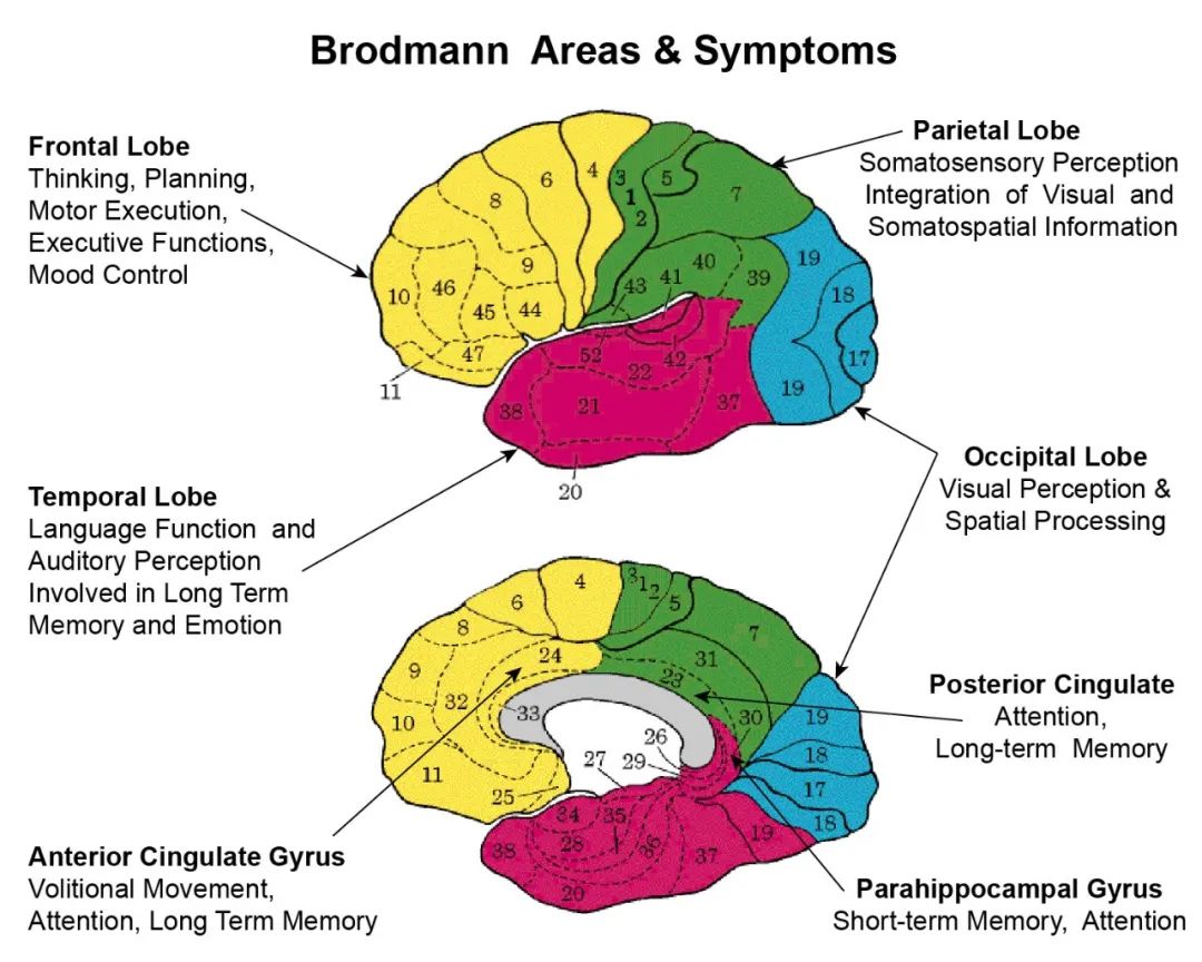
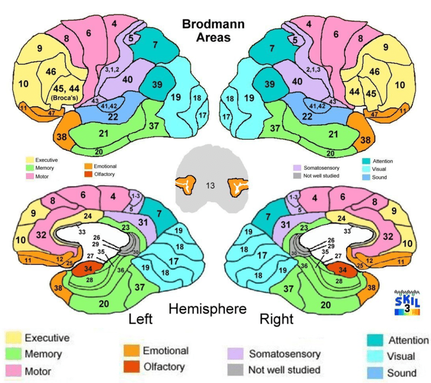
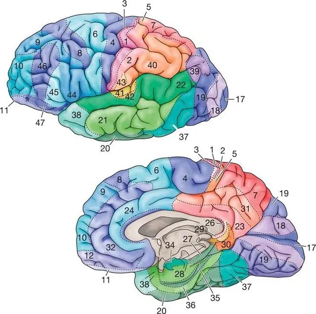
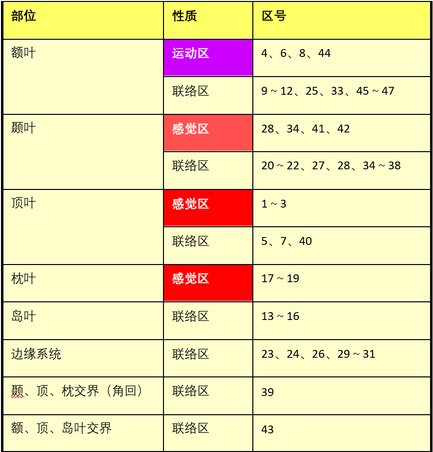 1, 2, and 3 Areas – Primary Somatosensory Cortex located in the postcentral gyrus and anterior parietal area. Function and Role: These areas are responsible for basic somatosensory functions, receiving sensory input from the contralateral limbs.4 Area – Primary Motor Cortex located in the precentral gyrus, medial side of the central sulcus. Function and Role: The primary motor cortex controls voluntary movements.5 Area – Somatosensory Association Cortex located in the anterior parietal cortex (the parietal cortex is part of the lower edge cortex). Function and Role: Forms the somatosensory association cortex with area 7.6 Area – Premotor Cortex and Supplementary Motor Cortex (Secondary Motor Cortex) located in the frontal lobe, anterior part of the precentral gyrus, is the premotor cortex and supplementary motor area. Function and Role: Together with area 8, it forms the premotor cortex, guiding sensory-motor coordination and assisting in the control of proximal and trunk muscles.7 Area – Somatosensory Association Cortex located at the top of the parietal cortex, posterior to the somatosensory cortex, above the visual cortex. Function and Role: Integrates visual and motor information, forming the somatosensory association cortex with area 5, responsible for visual-motor coordination.8 Area – Includes Frontal Eye Fields located in the frontal lobe, includes the frontal eye field. Function and Role: Together with area 6, it forms the premotor cortex, controlling voluntary eye movements, especially related to tracking movements.9 Area – Dorsolateral Prefrontal Cortex located in the frontal lobe, posterior lateral prefrontal cortex (involved in motor planning and organization).10 Area – Anterior Prefrontal Cortex (most rostral part of superior and middle frontal gyri) located in the frontal lobe, at the top of the inferior frontal gyrus (the most anterior part of the superior and middle frontal gyri) (involved in memory retrieval).11 Area – Orbitofrontal Area (orbital and rectus gyri, plus part of the rostral part of the superior frontal gyrus) located in the medial ventral surface of the frontal lobe, anterior frontal cortex area (orbital gyrus, rectus gyrus, and part of the anterior superior frontal gyrus). Areas 9, 10, and 11 together form the prefrontal cortex. Function and Role: Execute cognitive functions, encompassing all aspects of thought and perception, memory and recall of information, problem-solving, and emotions, closely linked to the limbic part of the forebrain.12 Area – Orbitofrontal Area (previously part of BA11, refers to the area between the superior frontal gyrus and the inferior rostral sulcus) located between the superior frontal gyrus and the inferior rostral sulcus (orbital area).13, 14 Areas – Insular Cortex15 Area – Anterior Temporal Lobe16 Area – Insular Cortex located in the insula. Areas 13-16 Function and Role: Process sensory experiences generated from converging information (such as feelings of disgust and anxiety), related to emotions. The anterior insula is involved in olfactory, gustatory, and limbic functions, while the posterior insula is associated with auditory, autonomic sensory-motor functions, and addiction.17 Area – Primary Visual Cortex (V1) located in the medial aspect of the occipital lobe (primary visual cortex, striate cortex V1), the main visual area. Function and Role: The initial site for processing visual information, organized into orientation columns and ocular dominance columns, receiving information from the lateral geniculate nucleus (below the posterior lateral thalamus) and sending information to other visual areas along dorsal and ventral pathways.18 Area – Secondary Visual Cortex (V2) located in the occipital lobe, including parts of the cuneus, lingual gyrus, and lateral occipital gyrus, part of the extrastriate cortex (secondary visual cortex, V2, extrastriate visual cortex), visual association area. Function and Role: Visual processing (visual association cortex).19 Area – Associative Visual Cortex (V3, V4, V5) located in the occipital lobe, visual association area. Function and Role: Visual processing, forming the visual association cortex with area 18.20 Area – Inferior Temporal Gyrus located in the inferior temporal gyrus. Function and Role: Higher-level information processing, visual ventral pathway. Sensory area – dorsal-ventral pathway. The ventral pathway enters the lower part of the temporal lobe (areas 20, 37), responsible for recognizing objects, text, and faces, i.e., “what,” while the dorsal pathway extends to the postcentral gyrus (areas 39, 40), perceiving spatial relationships, i.e., “where.”21 Area – Middle Temporal Gyrus located in the lateral temporal lobe, above area 20, below areas 40 and 41, middle temporal gyrus area. Function and Role: Language and auditory processing.22 Area – Superior Temporal Gyrus, of which the caudal part is usually considered to contain Wernicke’s Area located in the temporal lobe, its anterior part belongs to Wernicke’s area, auditory association area: judging the type of sound. Function and Role: The posterior part contains Wernicke’s area (including the superior temporal gyrus, posterior part of the middle temporal gyrus, supramarginal gyrus, and angular gyrus). It is the brain’s writing center and visual language center. Damage to Wernicke’s area will result in severe sensory aphasia, responsible for language comprehension.23 Area – Ventral Posterior Cingulate Cortex located in the occipital lobe, posterior end of the cingulate cortex, extending to the top of the parieto-occipital sulcus. Function and Role: As part of the limbic system, connected to the amygdala, orbitofrontal cortex, and hippocampus, involved in the emotional system.24 Area – Ventral Anterior Cingulate Cortex located in the ventral part of the cingulate cortex. Function and Role: As part of the limbic system, connected to the amygdala, orbitofrontal cortex, and hippocampus, involved in the emotional system.25 Area – Subgenual Area (part of the Ventromedial Prefrontal Cortex) located in the subgenual cortex. Function and Role: Related to olfaction.26 Area – Ectosplenial Portion of the Retrosplenial Region of the Cerebral Cortex located in the cingulate gyrus “gorge” area. Function and Role: Part of the memory system, responding to incidental events.29 Area – Retrosplenial Cingulate Cortex located in the retrosplenial cingulate cortex. Function and Role: Part of the memory system, responding to incidental events.30 Area – Subgranular Area, part of the cingulate gyrus. Function and Role: Part of the memory system, responding to incidental events.27 Area – Piriform Cortex located in the piriform cortex of the parahippocampal gyrus. Function and Role: Part of the memory system.28 Area – Ventral Entorhinal Cortex located in the posterior entorhinal cortex.31 Area – Dorsal Posterior Cingulate Cortex located in the dorsal posterior cingulate cortex. Function and Role: Emotion processing and recognition.32 Area – Dorsal Anterior Cingulate Cortex located in the parietal lobe. Function and Role: Responsible for the decision-making process.33 Area – Part of Anterior Cingulate Cortex part of the anterior cingulate cortex.34 Area – Dorsal Entorhinal Cortex (on the Parahippocampal Gyrus) part of the anterior cingulate cortex, located in the parahippocampal gyrus.35 Area – Perirhinal Cortex (in the rhinal sulcus) located in the parahippocampal gyrus.36 Area – Ectorhinal Area, now part of the perirhinal cortex (in the rhinal sulcus) located in the parahippocampal gyrus.The limbic lobe consists of the cingulate gyrus, parahippocampal gyrus, hippocampal gyrus, and its deep structures, forming a “C” shape around the corpus callosum, with functions related to the regulation of visceral activities; the limbic lobe and subcortical nuclei related to visceral activities, such as the hypothalamus, anterior thalamic nucleus, amygdala, midbrain tegmentum structures, and septal nuclei.37 Area – Fusiform Gyrus located in the temporal lobe, between the inferior temporal sulcus and the lateral sulcus. Function and Role: Multimodal integration, high-level object and face recognition.38 Area – Temporopolar Area (most rostral part of the superior and middle temporal gyri) located in the temporal pole, the anterior part of the superior and middle temporal gyri. Function and Role: Related to memory and emotions.39 Area – Angular Gyrus, considered by some to be part of Wernicke’s area, located in the parietal lobe. Function and Role: Semantic processing.40 Area – Supramarginal Gyrus, considered by some to be part of Wernicke’s area, located at the upper end of the lateral sulcus. Function and Role: Secondary somatosensory cortex, responding to bodily stimuli, completing structural differentiation tasks.41 Area – Auditory Cortex located in the temporal lobe, in the anterior transverse temporal gyrus of the medial aspect of the lateral sulcus. Function and Role: Early auditory information processing, low frequencies concentrated in the rostral side, high frequencies concentrated in the caudal/medial side.42 Area – Auditory Cortex located in the temporal lobe, at the edge of the lateral sulcus. Function and Role: Early auditory information processing, low frequencies concentrated in the rostral side, high frequencies concentrated in the caudal/medial side.43 Area – Primary Gustatory Cortex located in the subcentral area. Function and Role: Primary taste area.44 Area – Pars Opercularis, part of the inferior frontal gyrus and part of Broca’s area, located in the inferior frontal gyrus. Function and Role: Language motor area, related to the production of speech.45 Area – Pars Triangularis, part of the inferior frontal gyrus and part of Broca’s area, located in the frontal lobe, triangular area of the inferior frontal gyrus, above the upper boundary of the lateral sulcus. Function and Role: Language motor area, executing semantic tasks and producing written language.46 Area – Dorsolateral Prefrontal Cortex located in the dorsolateral prefrontal cortex, situated in the middle frontal gyrus of the frontal lobe, also part of the prefrontal cortex. Function and Role: Executive functions.47 Area – Pars Orbitalis, part of the inferior frontal gyrus, located in the frontal lobe, on the surface of the orbital gyrus. Function and Role: Processes grammar in language.48 Area – Retrosubicular Area (a small part of the medial surface of the temporal lobe).49 Area – Parasubicular Area in a Rodent located at the junction of the temporal lobe and insula.52 Area – Parainsular Area (at the junction of the temporal lobe and the insula).(*) Areas found only in non-human primates.Source: Movement and Rehabilitation Edited by: 3 Physical Therapists
Selected Past Articles
Popular Articles
Practical Physical Examination of the Ankle Joint
丨More
Discussion
How should tennis elbow be treated?
丨More
Discussion
Related Issues in Sports Injury Rehabilitation Practice
丨More
Discussion
Recommended Purchases


Add the editor’s WeChat: Oh-Physiotherapist Join group, submit, cooperate, consult

Disclaimer: Some images and texts are sourced from the internet for educational purposes, all rights belong to the original authors. If there is any infringement, please contact for removal. The content only represents the author’s personal views, and we hope everyone can judge and apply it rationally.