Invisible and intangible, yet capable of causing diseases in plants, animals, and humans, and rapidly spreading to trigger terrifying epidemics, do you know what viruses look like?
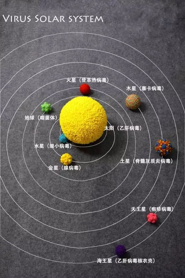
A solar system composed of virus models.
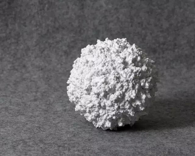
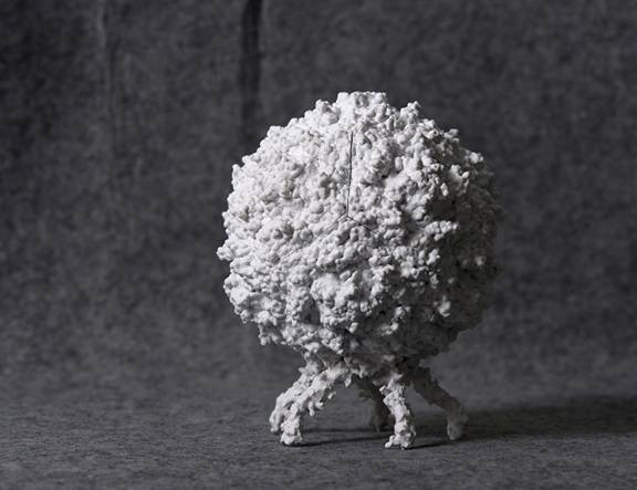
Utilizing the latest 3D printing technology, the Wuhan Institute of Virology, supported by the Chinese Academy of Sciences’ popular science project, has created several representative virus models based on research achievements in viral structural biology. These models restore the true appearance of virus structures, providing the most intuitive and vivid materials for understanding viruses. Let’s appreciate the masterpieces of nature and designers together!
Discovery of Viruses
The first virus discovered by humans was the Tobacco Mosaic Virus (TMV), which causes the tobacco mosaic disease and is a single-stranded RNA virus. In 1886, a German scientist named Mayer, working in the Netherlands, crushed the leaves of tobacco plants infected with mosaic disease, mixed them with water, and injected the juice into the veins of healthy tobacco leaves, causing them to develop mosaic disease, proving that this disease is contagious.
Leaves infected with the Tobacco Mosaic Virus and negative staining electron microscope images of the virus.
Through analysis of leaves and soil, Mayer pointed out that the tobacco mosaic disease is caused by bacteria. In 1892, Russian scientist Ivanovsky repeated Mayer’s experiment and further discovered that the juice from diseased tobacco leaves could still cause healthy tobacco plants to develop mosaic disease after passing through a bacterial filter, but Ivanovsky explained it as being due to toxins produced by bacteria. In 1898, Dutch bacteriologist Beijerinck also confirmed Mayer’s observations. He placed the juice from tobacco mosaic disease plants on the surface of an agar gel block and found that the infectious substance spread through the gel at a moderate rate, while bacteria remained on the surface of the agar. Beijerinck pointed out that the pathogenic factor causing tobacco mosaic disease has three characteristics: 1. It can pass through bacterial filters; 2. It can only reproduce within infected cells; 3. It cannot grow in non-living substances outside the host. Based on these characteristics, he proposed that this pathogenic factor is not a bacterium but a new substance, which he named “virus” from the Latin word for poison. The miraculous virus was “born”! Almost simultaneously, German bacteriologists Loeffler and Frosch discovered that the pathogen causing foot-and-mouth disease in cattle could also pass through bacterial filters, further confirming the significant discoveries of Ivanovsky and Beijerinck.
Representative Virus Models
1. Tobacco Mosaic Virus (TMV)
The Tobacco Mosaic Virus is rod-shaped, approximately 300 nanometers long and 18 nanometers in diameter, with its nucleocapsid protein arranged in a helical structure, and its nucleic acid is single-stranded RNA. TMV can infect over 310 species of plants across 30 families, especially tobacco and other solanaceous plants, causing diseases such as tobacco mosaic disease, with symptoms of mosaic patterns on the leaves, poor growth, and leaf deformities. TMV is widely distributed worldwide, with reports of its presence in Shandong, Hebei, Shanxi, Sichuan, Beijing, Shanghai, and other regions in China. TMV spreads through the rubbing of infected seedlings against healthy ones or through agricultural operations; additionally, insects with chewing mouthparts, such as locusts and tobacco cutworms, can also transmit TMV.
The 3D model showcases part of the structure of the Tobacco Mosaic Virus, with blue representing the viral coat protein and purple representing the genetic material RNA, exhibiting a regular helical arrangement and rod-like shape.
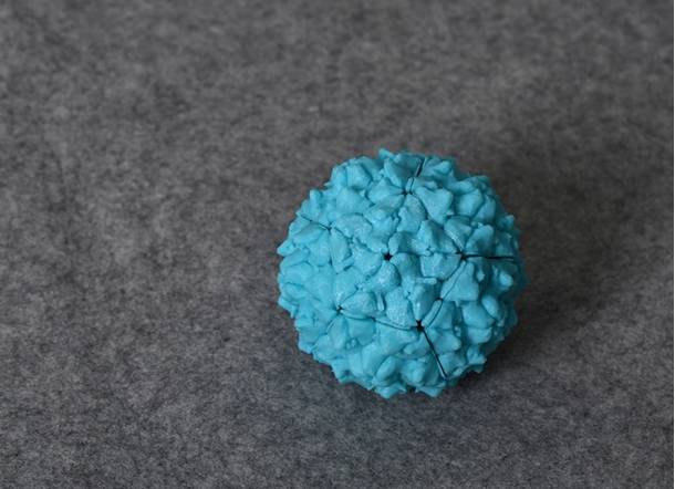
2. Poliovirus
The Poliovirus commonly invades the central nervous system, damaging the anterior horn motor neurons of the spinal cord, leading to flaccid paralysis of limbs, primarily seen in children, hence also known as infantile paralysis. The sugar cubes we ate as children were actually the poliovirus vaccine. Due to widespread vaccination over the years, the incidence of poliovirus in China has been well controlled, with a significant reduction in cases.
The Poliovirus belongs to the family of small RNA viruses. It primarily spreads through the digestive tract. Under an electron microscope, it appears as small spherical particles, with a diameter of 20-30 nm, exhibiting icosahedral symmetry. The virus particle’s center contains single-stranded positive-sense RNA, surrounded by 32 capsid subunits forming the outer shell, and this virus has a naked nucleocapsid without an envelope.
Humans are the only natural hosts for the Poliovirus because there is a receptor on the surface of human cell membranes that has a specific affinity for the structural protein VP1 on the virus coat, allowing the virus to attach to the cells.
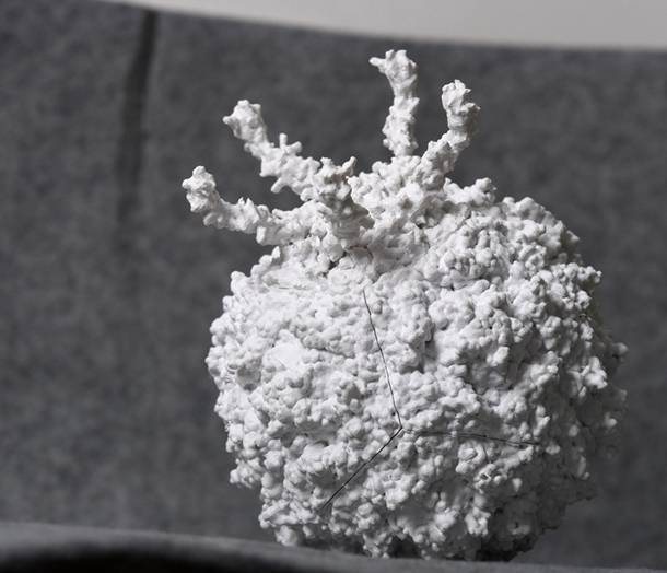
The long antennae in the image above are cell receptors that can bind to the structural proteins on the virus, facilitating viral infection of the cells.
At the same time, we simulated the self-assembly structure of the Poliovirus, and using magnets, we can assemble 12 independent virus structural protein components into a complete virus particle.
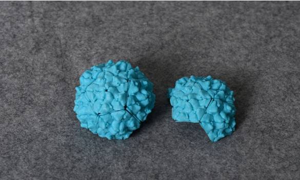

3. Hepatitis B Virus (HBV)
The Hepatitis B virus, abbreviated as HBV, is a DNA virus belonging to the Hepadnaviridae family. The infection rate of HBV in China is approximately 60%-70%; the carrier rate of Hepatitis B surface antigen (HBsAg) is about 7.18% of the total population, which translates to approximately 93 million people carrying the HBV, with about 20 million cases of chronic Hepatitis B.
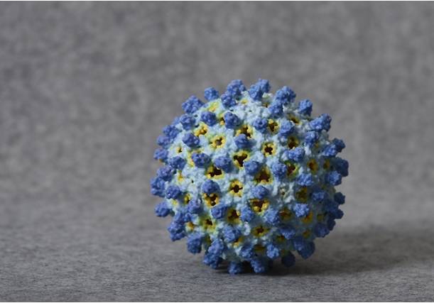
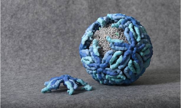
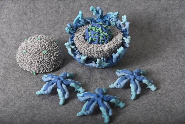
This image shows the nucleocapsid of the Hepatitis B virus, which is an icosahedron composed of core antigens.
The nucleocapsid is assembled from 12 subunits, each subunit is composed of 5 tetramers, and each tetramer is made up of 4 core antigen proteins, totaling 240 core antigens (HBcAg).
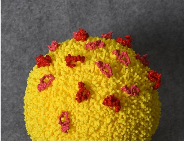
The outermost layer of the complete Hepatitis B virus consists of three types of Hepatitis B surface antigens (HBsAg, red) and an envelope (yellow).
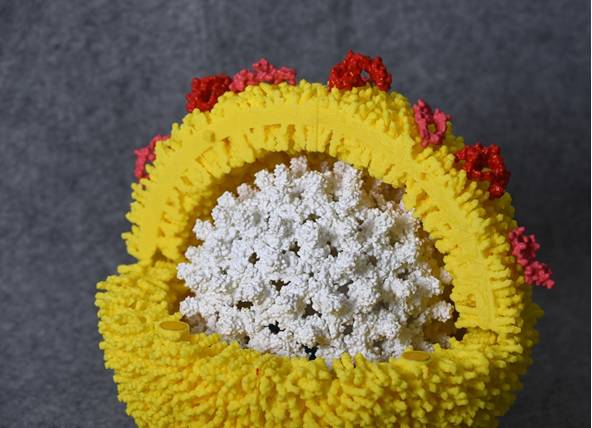
The envelope encloses the nucleocapsid assembled from core antigens (HBcAg, white).

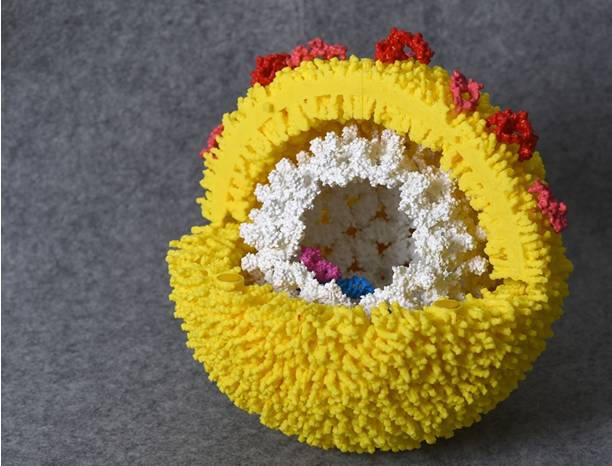
The nucleocapsid encloses the viral genetic material DNA (blue) and the DNA polymerase (purple) responsible for replication.
Hepatitis B Brief Science:
Hepatitis B virus (HBV) carriers are defined as patients who are HBsAg positive, meaning the red protein can be detected, but their liver function is normal.
Large three positives and small three positives are classified based on the differences in the patient’s HBV serological markers (the five items of Hepatitis B):
1. Large three positives refer to HBsAg+, HBeAg+, anti-HBc+, meaning the detection of red protein, similar to white protein (HBeAg and white protein are similar, more present when the virus is actively replicating), and the antibody to white protein.
2. Small three positives refer to HBsAg+, anti-HBe+, anti-HBc+, meaning the detection of red protein, the antibody similar to white protein, and the antibody to white protein.
It can be seen that the difference between the two is whether HBeAg or its antibody is positive. Generally, HBeAg+ indicates active viral replication, with a higher HBV DNA load, thus the HBV DNA detection in the patient’s bodily fluids is often positive, posing a higher risk of transmission to others. Note: + indicates positive.
HBsAg, anti-HBs, HBeAg, anti-HBe, and anti-HBc are the five items for Hepatitis B testing.
Anti-HBs and anti-HBc being positive generally indicates vaccination against Hepatitis B.
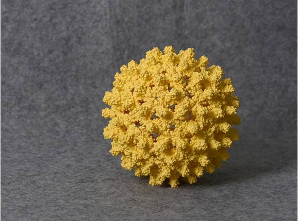
4. Zika Virus (ZIKV)
The Zika virus recently caused an outbreak in Brazil from 2015 to 2016, resulting in a large number of infants with microcephaly, causing global panic.
The incubation period for Zika virus disease (the time from exposure to the onset of symptoms) is not clearly defined but may be a few days. Symptoms of the disease are similar to those of other arthropod-borne viral infections such as dengue fever, including fever, rash, conjunctivitis, muscle and joint pain, general weakness, and headache. These symptoms are often mild and last for 2-7 days.
The Zika virus is transmitted to humans primarily through the bites of infected Aedes mosquitoes, mainly in tropical regions. Aedes mosquitoes typically bite during the day, with peak periods in the early morning and evening/night. These mosquitoes also transmit dengue fever, chikungunya, and yellow fever. Zika virus may also be sexually transmitted. Currently, investigations are underway into other transmission methods, such as through blood transfusions.
The Zika virus itself is not highly harmful; 80% of adult infections are asymptomatic, and 20% of patients may only exhibit mild cold-like symptoms. However, pregnant women infected with the Zika virus can transmit it to their infants, affecting brain development and causing microcephaly.
The Zika virus belongs to the Flavivirus family. The yellow fever virus was the first human virus discovered in history (the first virus discovered was the Tobacco Mosaic Virus), identified in 1900, confirmed to be transmitted by mosquitoes.
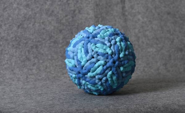
The Zika virus has a diameter of 50 nanometers, with the outer layer composed of assembled membrane protein E (blue), and the interior wrapped in a lipid membrane (gray).
The membrane also has small membrane protein M (cyan), and the core wrapped by the membrane consists of capsid proteins (green) and genomic RNA (dark blue).
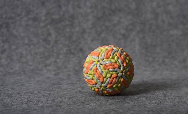
Virus Art
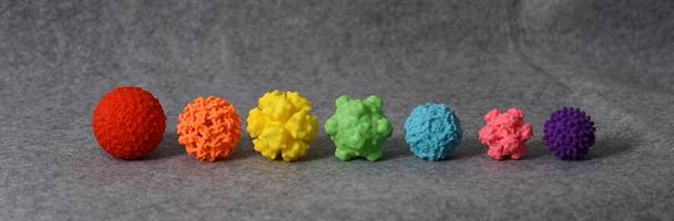
Dengue virus, West Nile virus, adenovirus, bacteriophage (head), parvovirus, herpesvirus, Hepatitis B virus (nucleocapsid).
The author is a researcher at the Wuhan Institute of Virology, Chinese Academy of Sciences
Source: Wuhan Institute of Virology, Chinese Academy of Sciences