Multifocal hepatic steatosis (also known as multifocal nodular hepatic steatosis) is a rare condition characterized by multiple focal fat deposits in the liver, which can sometimes be confused with liver metastases.
Epidemiology
Risk Factors: Similar to other forms of hepatic steatosis, including:
-
Diabetes
-
Obesity
-
Chronic excessive alcohol consumption
-
Exogenous steroids
-
Medications (such as amiodarone, methotrexate, chemotherapy drugs)
-
Parenteral nutrition
Clinical Manifestations
Multifocal hepatic steatosis is usually an incidental finding on imaging studies.
Pathology
The characteristic location is the medial segment of the left lobe of the liver (segment 4), located anterior to the porta hepatis or near the falciform ligament. It is believed to be related to variations in vascular supply.
Imaging Features
The size of the steatotic lesions ranges from a few millimeters to several centimeters, with no mass effect (i.e., they do not displace liver vessels or other structures), and no internal vascular enhancement.
Ultrasound
-
Focal steatosis typically appears as well-defined hyperechoic areas, with no mass effect.
-
May be accompanied by acoustic shadowing.
-
Color Doppler usually shows no blood flow or only minimal blood flow [1,2].
CT
-
Focal steatosis appears as low-density lesions, with no mass effect or significant enhancement.
MRI
The most distinguishing MRI feature is out-of-phase low signal.
Signal Characteristics:
-
T1WI: Lesions appear as high signal
-
T2WI: Mild signal increase
-
T1 enhancement (Gd-EOB-DTPA): During the hepatobiliary phase (20 minutes), the liver shows uniform enhancement, with the degree of enhancement of steatotic lesions consistent with normal liver tissue
-
In-phase/Out-of-phase (IP/OP):
-
In-phase: Steatotic lesions appear as high signal
-
Out-of-phase: Signal loss (fat suppression)
-
DWI/ADC: Usually no diffusion restriction
Case: A 48-year-old male, incidental finding of multiple focal liver lesions during a health check-up.
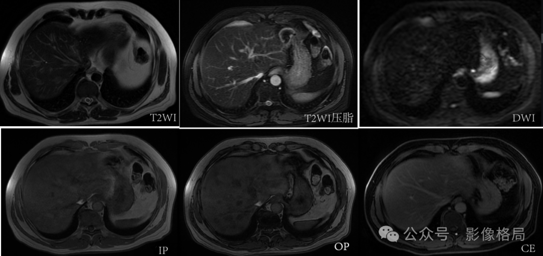
Multiple well-defined lesions in the liver parenchyma, high signal on T1 and T2, with reduced signal intensity on T2 fat-suppressed and out-of-phase sequences. No diffusion restriction on DWI/ADC sequences. No abnormal enhancement on contrast scans. — Multifocal hepatic steatosis.
A 65-year-old female, multiple low-density lesions in the liver.
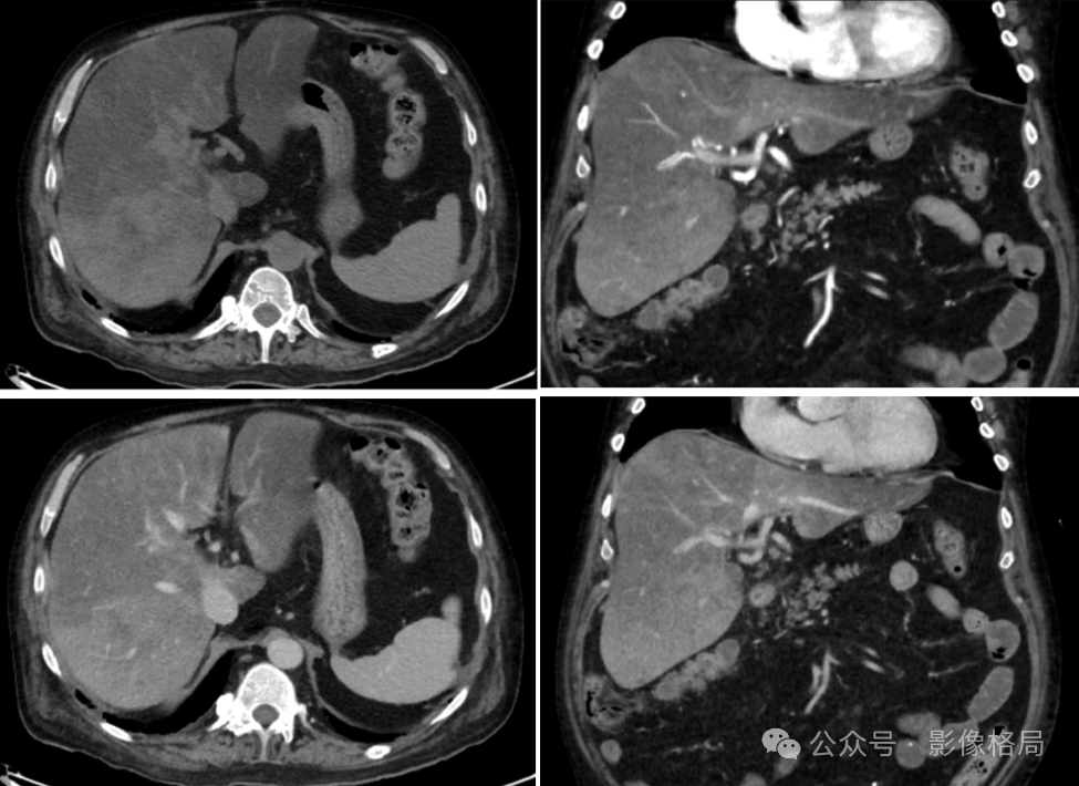
From left to right, top to bottom, are plain scan, arterial phase, venous phase, and delayed phase.
Heterogeneous signal in the liver, with a plain scan HU value of -4, indicating hepatic steatosis. No displacement or narrowing of vessels. No lymphadenopathy. No ascites.
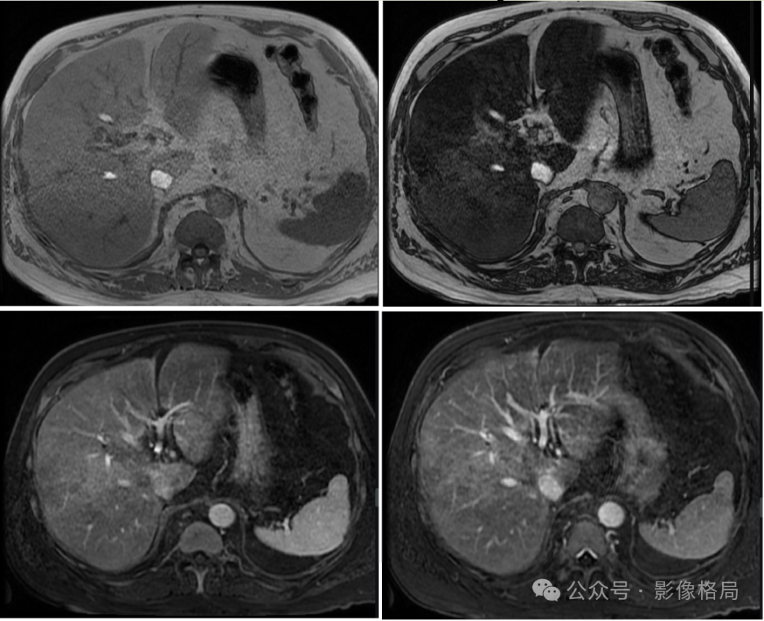
From left to right, top to bottom, are T1WI in-phase, T1WI out-of-phase, T1WI+C, and T1WI+C fat-suppressed.
The liver tissue shows diffuse signal reduction on out-of-phase, confirming nodular/map-like steatosis.