
The team led by Professor Young-Tae Chang at Pohang University of Science and Technology in South Korea has developed a fluorescence probe (SLY) based on boron-doped heteroaromatic compounds for the specific recognition of sialylated Lewis antigens (sLex and sLea) that are overexpressed on the cell surface, achieving high-fidelity fluorescence imaging of cancer regions at both cellular and tissue levels.
Cell surface glycosylation plays a crucial role in various biological processes, affecting cell communication, immune responses, and disease mechanisms. The composition and structure of cell surface glycans change with cell status, making them biomarkers for identifying cell types and states. Sialylated glycans are overexpressed in various cancers and are important targets for cancer diagnosis and treatment. However, existing methods (such as mass spectrometry) are difficult to implement for rapid detection and real-time imaging due to expensive equipment and complex procedures.
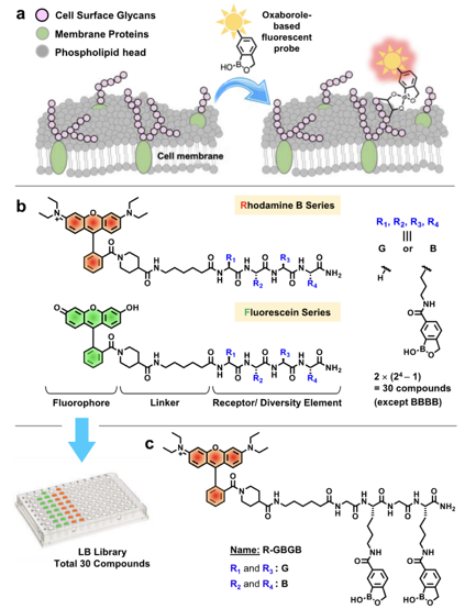
The researchers constructed a library of probes based on fluorescent boronic acid (LB) and systematically evaluated the structural factors affecting the probe’s ability to recognize and differentiate cell surface glycans. In the probe design, boron-doped heteroaromatic compounds serve as sugar-binding units, which, compared to traditional boronic acids, have a faster hydration rate and lower pKa at neutral pH, allowing for specific binding to sialic acid; for the fluorophore selection, Rhodamine B and fluorescein were used to assess their impact on probe binding, cellular localization, non-specific binding, and imaging capabilities; the linker chain consists of 6-aminohexanoic acid and 4-piperidine carboxylic acid, ensuring that the linker chain terminus is a secondary amine to prevent fluorescence quenching; the peptide backbone was synthesized using Fmoc solid-phase synthesis, randomizing the N-terminal amino acid residues and introducing boron-doped heteroaromatic-modified lysine to form a diverse probe library.
In vitro experiments showed that the SLY probe exhibited significant specific staining in HepG2 and HT29 cells, with fluorescence intensity over 10 times higher than the control group, while staining was weak in cells that do not express sLex and sLea; mechanistic studies revealed that SLY enters cells through caveolin-mediated endocytosis by binding to sialic acid on the cell surface, ultimately localizing to the mitochondria; in a liver cancer mouse model, SLY was able to distinguish normal and cancerous regions in frozen sections and fresh tissue samples, showing good co-localization with anti-Glypican-3 antibodies; after intravenous injection of SLY, the liver region of liver cancer model mice exhibited significant fluorescence signals, while normal mouse livers showed no fluorescence, validating SLY’s ability to label cancer tissues in vivo.
In summary, the SLY probe developed in this study provides a new tool for cell labeling, cancer diagnosis, and fluorescence-guided surgery, with its optimized recognition group quantity and spatial arrangement, as well as fluorophore selection, offering important references for future glycan-targeted probe design.
Original link:
https://pubs.acs.org/doi/10.1021/jacs.5c03020
Author: HYQ
Proofreader: LYP
Editor: LYP