
【Quote】
In Vitro Diagnostics (IVD) has become an indispensable tool for clinical diagnosis and monitoring of disease progression. Current IVD technologies mainly include molecular biology diagnostics, immunoassays, and physiological signal monitoring. Among these, immunoassays have been routinely used to detect various biomarkers such as small molecules, proteins, and nucleic acids. The Enzyme-Linked Immunosorbent Assay (ELISA) has become the most commonly used immunoassay method due to its good specificity, sensitivity, and high throughput. However, traditional ELISA methods not only require complex operational procedures and expensive instruments but also lack multiplexing and good reproducibility, limiting their application in resource-limited areas. Furthermore, for early diagnosis or screening of specific diseases such as Alzheimer’s disease and acute myocardial infarction, traditional ELISA is insufficient to detect at the nanomolar level. Therefore, it is particularly important to develop new types of ELISA with ultra-high sensitivity, good reproducibility, and multiplexing capabilities.
In the past two decades, various significant technological innovations have emerged in ELISA-derived technologies. For example, to develop low-cost, stable, and repeatable target-recognizing molecules, aptamers have been introduced into ELISA to replace traditional antibodies in various biosensor platforms. To enhance simplicity and achieve on-site detection, paper-based ELISA has been developed utilizing capillary action to drive liquid flow. To improve detection sensitivity, various amplification strategies (such as nucleic acid amplification and nanomaterials) have been employed in ELISA. To enhance multiplex detection capabilities, magnetic beads and microfluidic chips have been integrated into ELISA. Thanks to these innovations, emerging ELISA-derived technologies, such as aptamer-based ELISA, paper-based ELISA, digital ELISA, and plasmonic ELISA (Figure 1), have made significant progress and show great potential in IVD applications, especially in point-of-care (POC) settings.
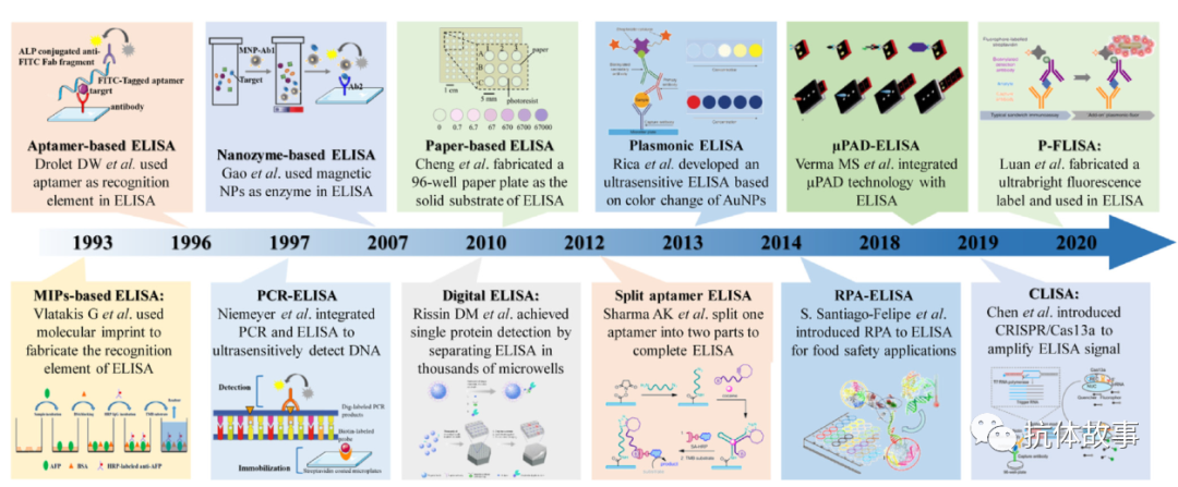
Figure 1. Overview of representative ELISA-derived technologies in recent decades
This article provides a clear and comprehensive overview of new ELISA-derived technologies and prospects, briefly summarizing the historical development of ELISA and outlining its recent directions, introducing various new ELISA (i.e., aptamer-based ELISA, digital ELISA, nanozyme ELISA, paper-based ELISA, plasmonic ELISA, CLISA, and plasmonic fluorescence-enhanced ELISA) and their applications in IVD. Finally, several promising development directions for ELISA-derived technologies are proposed, aiming to accelerate the development of ELISA-derived technologies and hoping to usher in a new era of ultra-sensitive and portable immunoassays through new ELISA-derived technologies.
【Content Introduction】
1. Development of ELISA
In 1959, Ylow and Berson combined the high sensitivity of radioactive isotope tracing technology with the high specificity of immunoassays to determine the insulin levels in the plasma of diabetic patients, thus pioneering radioimmunoassay. To avoid the health hazards posed by radioactive substances, in 1971, enzymes were used to replace radioactive materials, forming a new colorimetric/fluorescent immunoassay method, namely ELISA. The first use of alkaline phosphatase as a reporter marker to detect immunoglobulins in rabbit serum by measuring the corresponding absorbance featured biosafety and high throughput, leading to the rapid development of commercial ELISA kits widely used in research and diagnostics. To simplify operations and shorten detection time, fully automated ELISA systems were subsequently developed, enabling rapid, ultra-high throughput clinical diagnostics. Currently, ELISA has become a routine testing tool in the food industry, vaccine development, clinical diagnostics, toxicology, pharmaceutical industry, and organ transplantation. ELISA is based on the specific recognition between paired antigens and antibodies. ELISA typically consists of four main components: solid-phase carrier, recognition element, signal amplification part, and readout method. The properties of these components largely determine the performance of ELISA, and all new ELISA-derived technologies have improved at least one of these four parts.
ELISA is a heterogeneous immunoassay method that usually occurs on the surface of a solid-phase carrier. The solid-phase carrier is used to immobilize capture antibodies and separate reactants from non-reactants. Common solid-phase carriers include microtiter plates, MBs (methyl methacrylate, butadiene, styrene terpolymer), and paper materials. Microtiter plates are made from low-cost, transparent, easily modified, low-background signal, and protein-friendly materials such as polystyrene (PS), polymethyl methacrylate (PMMA), and polycarbonate (PC). Due to the integration of multiple wells (i.e., 48, 96, 384), microtiter plates can be used for parallel analysis of multiple samples. MBs with high specific surface area can adsorb a large amount of antibodies and expose epitopes fully to target antigens. At the same time, MBs facilitate the separation of complexes, avoiding multiple washing steps in the ELISA method. Therefore, compared to microtiter plate ELISA, MB-based ELISA is more sensitive and easier to automate, but its surface may induce non-specific binding of proteins and antibodies, leading to potential detection errors. The immobilization of antibodies on paper materials is an emerging technology that mainly utilizes the porous structure and capillary action of paper materials to achieve passive reagent delivery. It reduces reliance on instruments and production costs, making it particularly suitable for POC diagnostics. Additionally, in fewer cases, small tubes larger than microtiter wells have also been used to immobilize antibodies for ELISA. Thus, sensitivity can be improved by increasing the amount of samples and reagents. Small tubes can also be used, which are then directly placed into a spectrophotometer to compare colorimetric signals. Although tube ELISA is a cost-effective method, it has a lower sample throughput. To address this issue, a capillary array-based ELISA similar to tube ELISA has been developed, which can significantly increase sample throughput and reduce detection time compared to microtiter plate-based ELISA.
The main recognition element in traditional ELISA is antibodies. The affinity of antibodies for target antigens is a core factor determining the performance of ELISA. Antibodies are produced by the immune systems of animals (e.g., rabbits, mice, goats) to recognize and neutralize foreign substances. Among them, mice are the most studied animal model for producing various monoclonal and polyclonal antibodies, and there are now many available antibodies against mouse genes. Therefore, mouse antibodies have been widely used in early detection research, in vivo preclinical therapeutic studies, IVD, and other fields. In contrast, rabbit antibodies are relatively new, with higher affinity and specificity compared to mouse antibodies. Rabbit antibodies are more closely related to human antibodies and can recognize a wider variety of epitopes. Additionally, when large amounts of antibodies are needed, goats are usually preferred, as they can produce 7 to 8 times more antiserum than other small animals, resulting in antibodies that are 2 to 3 times richer than those from other animals. Therefore, the choice of antibodies depends on the research purpose. The preparation of commercial antibodies mainly uses hybridoma technology. Additionally, phage display technology and genetic engineering are utilized to screen for high-affinity antibodies. The specificity and affinity of antibodies also highly depend on the immunogen designed based on the target antigen. Therefore, reasonable design of the immunogen is beneficial for obtaining the desired high-affinity antibodies, thereby improving the sensitivity of ELISA.
The signal amplification part in conventional ELISA refers to enzymes, which are generally combined with secondary antibodies. Commonly used enzymes include horseradish peroxidase (HRP), alkaline phosphatase (ALP), and β-D-galactosidase (GAL), among which HRP is the most widely used due to its small size and fewer spatial issues. However, HRP is sensitive to preservatives such as sodium azide and metal ions. Additionally, during the catalytic process mediated by HRP, the reactant H2O2 can also inhibit the catalytic reaction, thus limiting the effective time for substrate incubation. ALP is much more expensive than HRP, but it has many advantages in immunoassays. For example, it has high thermal stability and catalytic efficiency, but it may be inhibited by metal chelators (such as cysteine, ethylenediaminetetraacetic acid, thioethanol). GAL’s most commonly used chromogenic substrate is o-nitrophenyl-β-D-galactopyranoside (ONPG), which has a relative catalytic rate of up to 500 U/mg and minimal solid-phase interference. Therefore, galactosidase has broad prospects in developing very sensitive and simple protein homogeneous immunoassays. Depending on the type of substrate, the generated signals can be colorimetric, fluorescent, or chemiluminescent. Generally, ELISAs with fluorescent or chemiluminescent signals can achieve higher detection sensitivity.
The reading of traditional ELISA results relies on the naked eye or microplate readers. Naked-eye readings based on colorimetric signal analysis are expected to enable high-throughput screening tests and POC diagnostics in resource-limited areas, but they only provide qualitative results. Most automated microplate readers are equipped with unique software programs to read absorbance or fluorescence results, enabling rapid quantitative readings. However, microplate readers are large and often limit the application of POC. Recently, some smartphone-based microplate readers have been developed, reducing the size of the instruments and making on-site detection possible.
To continuously improve the performance of ELISA, researchers have proposed strategies to optimize these components (Figure 2), resulting in various new ELISA-derived technologies, such as MIP-based ELISA, aptamer-based ELISA cards, PCR-ELISA, paper-based ELISA, digital ELISA, plasmonic ELISA, and plasmonic fluorescence-enhanced FIISA (P-FLISA). To better understand these strategies, this article reviews these technologies and their applications in IVD.
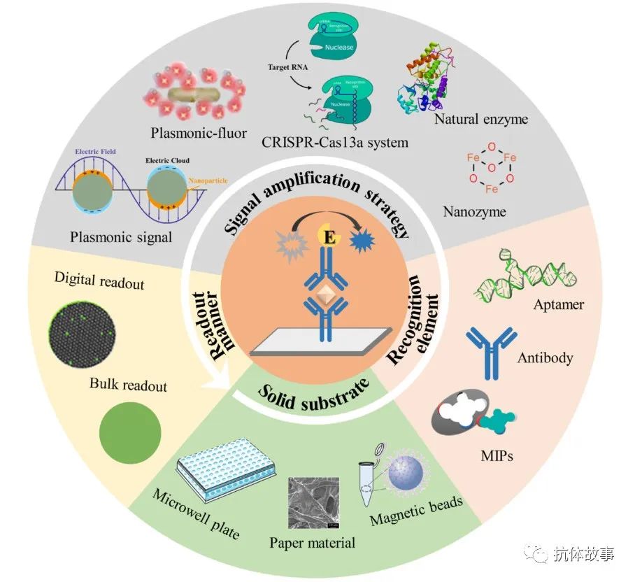
Figure 2. Summary of improvements in ELISA-derived technologies
2. Innovative New ELISA-Derived Technologies Based on Solid-Phase Carriers
The solid-phase carrier captures antibodies in ELISA. Currently, three types of solid-phase carriers (i.e., microtiter plates, paper materials, and MBs) are used for ELISA, forming three different types of ELISA-derived technologies, each with its advantages and disadvantages (Figure 3).
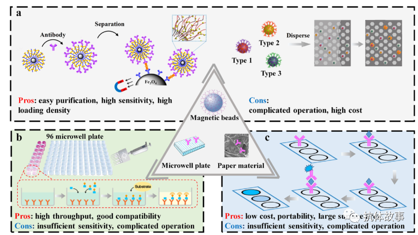
Figure 3. Comparison of main solid-phase carriers in ELISA
2.1. Microtiter Plate ELISA
Microtiter plate ELISA is the most commonly used method, which is sensitive but complex to operate, including microtiter plate coating and blocking, multiple washing and incubation steps, and colorimetric reading. In recent years, many studies have focused on optimizing various steps of microtiter plate ELISA to improve its sensitivity and specificity. Blocking is a very important step in ELISA as it relates to non-specific binding and detection specificity. Improper blocking treatment may lead to high background signals and low sensitivity. Common blocking agents include bovine serum albumin (BSA), non-fat dry milk, fetal bovine serum, or whole serum. However, due to the steric hindrance of the capture antibodies, the coated macromolecular layer may inhibit the binding efficiency of the capture antibodies. Therefore, some polymer molecules have been used to block ELISA microtiter plates. Microtiter plate ELISA requires washing, but the operation is complex. To simplify the microtiter plate-based ELISA procedure, most laboratories have replaced manual washing with automated plate washers. In terms of reading, chemiluminescence and fluorescence reading methods have also been adopted to improve detection sensitivity. The 96-well microtiter plate is the main form of ELISA established on the basis of microtiter plates. To process more samples in parallel and/or reduce the consumption of samples/reagents, 384-well regular volume plates and 384-well low-volume plates are also used in practical analysis. Although microtiter plate ELISA analyzes a single analyte using specific antibody pairs, the emergence of proteomics has led to the demand for multi-component analysis, and the cross-reactivity of antibodies is the main challenge in achieving multi-component detection in microtiter plate ELISA.
2.2. Paper-Based ELISA
Even with high sensitivity, microtiter plate ELISA still requires laboratory environments and takes several hours to complete. This time-consuming and complex ELISA method cannot meet the demands for rapid high-throughput diagnostics and POC testing. Therefore, there is an urgent need to develop new ELISA-derived technologies that have short analysis times and on-site detection capabilities. Recently developed paper-based diagnostic platforms have achieved rapid, inexpensive, and simple detection, showing good prospects.
Paper is widely used in scientific research and industrial applications due to its availability, low cost, light weight, and portability. Paper-based biosensors are reliable platforms for detecting various targets. The porous structure of paper materials has strong capillary action, making it a self-driven platform and exposing more surface area to immobilize recognition biomolecules. However, effectively coating antibodies on paper and preventing their desorption from the paper material poses certain challenges. To improve the coating effect, various methods have been proposed, such as chitosan-glutaraldehyde crosslinking, physical adsorption, and biotin-avidin systems, but most of these methods have drawbacks such as long coating times and poor coating effects.
Paper-based ELISA combines the sensitivity and specificity of ELISA with the simplicity, low cost, and user-friendliness of paper solid-phase carriers. Therefore, it is faster and cheaper than traditional ELISA. Additionally, it can be read using inexpensive desktop scanners or mobile cameras, minimizing equipment requirements and enabling disease diagnosis in resource-limited areas. However, compared to traditional ELISA, the sensitivity of paper-based ELISA is reduced. Therefore, it is necessary to further improve the sensitivity of paper-based ELISA. Another important direction is to develop integrated and automated paper-based ELISA for all sample processing steps.
2.3. Magnetic Bead ELISA
A disadvantage of microtiter plate ELISA is the steric hindrance and small specific surface area of the microtiter plate solid-phase matrix, leading to long reaction times. In ELISA, MBs are used as freely movable solid substrates, achieving a larger specific surface area and higher reaction activity by accelerating the diffusion process. Additionally, MBs can utilize magnetic force to simply and quickly separate the components of interest from the analytical system. The aforementioned characteristics of MBs endow MB-based ELISA with rapid reactions and high sensitivity.
Replacing microtiter plates with MBs indeed improves the detection sensitivity of ELISA and shortens reaction times. However, their dependence on specific magnetic devices or equipment increases costs. Additionally, the loss of MBs during washing may also affect the accuracy of detection.
3. Innovative New ELISA-Derived Technologies Based on Recognition Elements
Antibodies are the most widely used recognition elements in conventional ELISA. Recently, nucleic acid molecules and molecularly imprinted polymers (MIPs) have been used in ELISA-derived technologies, showing different advantages in terms of cost, stability, specificity, and affinity (Figure 4).
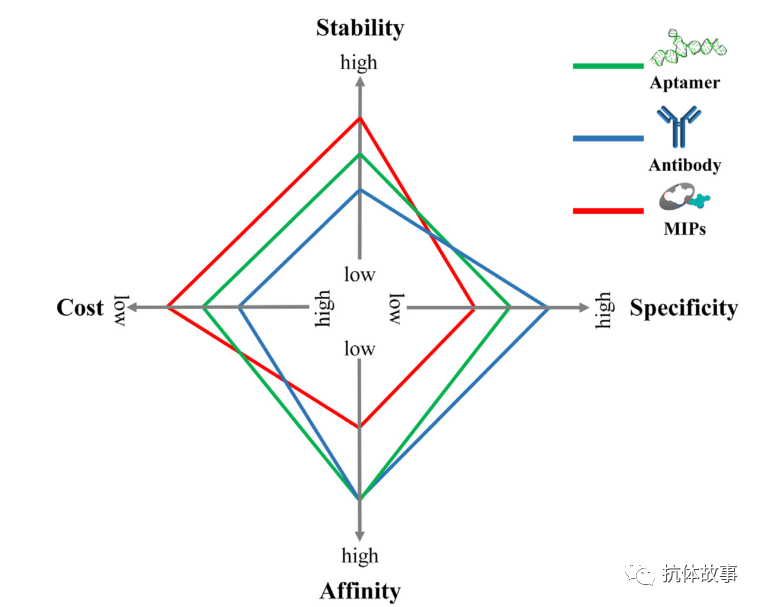
Figure 4. Comparison of main recognition elements in ELISA-derived technologies in terms of cost, stability, specificity, and affinity
3.1. Aptamer-Based ELISA
Traditional ELISA uses antibodies as recognition molecules, which have limitations such as poor stability, high costs, inevitable batch differences, and difficulty in large-scale production. Moreover, for certain targets, such as nucleic acids and non-immunogenic small molecules, it is also challenging to find corresponding antibodies, which limits its application. To address these issues, aptamers have been introduced into ELISA due to their advantages of good reproducibility, low cost, and wide target recognition range. Aptamers are short single-stranded nucleic acid sequences that can recognize and bind to various molecules. In 1997, Drolet et al. first used oligonucleotide aptamers as recognition elements in ELISA, resulting in a method called enzyme-linked aptamer sorbent assay (ELASA) or aptamer-based ELISA. Since then, various forms of aptamer-based ELISA have been developed, among which competitive and sandwich aptamer-based ELISA are the most representative.
Competitive aptamer-based ELISA is performed by having the target compete with labeled targets or mimic molecules for binding to the aptamer, and the released labeled molecules can induce subsequent signal generation. Competitive aptamer-based ELISA is suitable for small molecule detection, but it is generally difficult to find matching recognition elements. For sandwich aptamer-based ELISA, various types have been developed, such as aptamer-target-aptamer, aptamer-target-antibody, and antibody-target-aptamer. The aptamer-target-aptamer method requires a pair of aptamers to bind to the target at different binding sites, forming a sandwich structure. This sandwich method is suitable for macromolecules such as proteins that have sufficient binding sites. Since capture aptamers are easy to reuse and detection aptamers are easy to label, the aptamer-based sandwich ELISA method provides a significantly cost-effective approach compared to antibody-based sandwich ELISA. Another type of sandwich aptamer-based ELISA simultaneously utilizes antibodies and aptamers. However, the sandwich aptamer-based ELISA method is not suitable for small molecules, as most small molecules do not have two or more binding sites.
Currently, aptamer-based ELISA is limited to scientific research, as the overall success rate of aptamer selection still needs improvement; the binding ability of aptamers is highly related to their three-dimensional structure, which is influenced by various factors such as incubation conditions and buffer composition. With further development of aptamer technology, aptamer-based ELISA may find broader applications.
3.2. Molecularly Imprinted Polymer-Based ELISA
In addition to aptamers, another technology has been developed to obtain a low-cost, stable, and reproducible recognition element for ELISA, namely MIPs or “plastic antibodies.” MIPs are chemically synthesized receptors with antibody-like binding properties, formed by crosslinking polymerization of functional monomers around template molecules. Compared to antibodies, MIPs have advantages such as short synthesis cycles, low costs, and good environmental stability, making them considered promising alternatives to antibodies.
The first MIP-based ELISA was based on the binding analysis of competitive radioactive-labeled ligands. Similar to solid-phase radioimmunoassay, MIPs replaced the coated antibodies, leading to the development of MIPs-based ELISA. Among them, MIPs-coated 96-well microtiter plates are the most commonly used type and have made significant progress in recent years.
Common biomarkers in IVD are peptides and proteins. Although molecular imprinting technology has been widely developed, efficient imprinting of biomolecules such as proteins remains very challenging, making it difficult to achieve precise control over the imprinting process for biomolecules of different sizes, and the specificity of the prepared MIPs still needs further improvement. The combination of MIPs with nanomaterials avoids dependence on enzymes or antibodies in various assays and can be easily applied to existing analytical formats. It is believed that MIPs-based ELISA will overcome current limitations and achieve unprecedented development in the near future.
4.3. PCR-ELISA
In addition to newly emerging antigen recognition elements, nucleic acids have also been used to detect PCR products. PCR is a conventional gene amplification technique. However, its traditional readout methods can only provide qualitative results or pose certain safety hazards (i.e., gel electrophoresis and radioactive isotope labeling). In 1997, Nimeyer et al. first established the PCR-ELISA method, which utilizes ELISA as a readout method for PCR products. A typical method involves denaturing the PCR products with labeled digoxin (DIG), hybridizing the PCR products with capture probes on microtiter plates, adding enzyme-labeled antibodies against DIG, and finally adding chromogenic substrates to measure absorbance with an ELISA plate reader. Although PCR-ELISA is cumbersome and costly, it only requires a thermal cycler and microplate reader for quantifying PCR products.
PCR-ELISA inherits the high sensitivity and specificity of PCR technology and the batch detection capability of ELISA. The sensitivity of this method is about 100 times that of agarose gel electrophoresis. Therefore, this method is widely used for the detection of bacteria, fungi, parasites, viruses, and telomerase. Using basic laboratory equipment, quantitative and qualitative analyses can be performed in a short time. However, PCR-ELISA cannot detect unknown genes, as it requires specific primers and probes to amplify the target gene. Additionally, compared to real-time fluorescent polymerase chain reaction, PCR-ELISA can only provide the quantity of nucleic acid amplification fragments at the endpoint, losing quantitative information during the amplification process. PCR-ELISA successfully applies the principles of ELISA to the detection of high-throughput, low-cost polymerase chain reaction products, making it a very powerful analytical tool.
5. Innovative New ELISA-Derived Technologies Based on Signal Amplification
Signal amplification is an important aspect related to the sensitivity of ELISA. Traditional ELISA uses enzyme-catalyzed substrates to generate colorimetric signals. Many studies have focused on introducing new signal amplification strategies into ELISA, such as nanozymes, plasmonic signals, CRISPR-Cas systems, and enhanced labels (Figure 5).
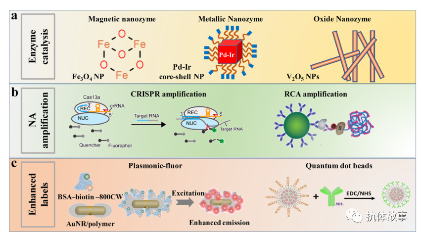
Figure 5. Comparison of major signal amplification strategies in ELISA-derived technologies
5.1. Nanozyme-Based ELISA
Traditional ELISA relies on natural enzymes such as HRP and ALP for signal amplification. Although these enzymes have high substrate specificity and catalytic efficiency, their application is still limited by some inherent drawbacks of natural enzymes, such as high preparation/purification costs, non-recyclability, and low catalytic activity under extreme pH and high-temperature conditions. In recent years, many nanoparticles with strong catalytic activity have been discovered, referred to as nanozymes, and introduced into ELISA, resulting in nanozyme-based ELISA, with researchers modifying them through a series of studies.
Nanozymes have many advantages, including low cost, high stability, and ease of surface modification, leading to significant development in the field of immunoassays. However, they still have certain limitations that hinder their replacement of natural enzymes as conventional ELISA labels. Although many nanomaterials have been proven to be enzyme mimetics, their catalytic activity is still far below that of corresponding natural enzymes. Furthermore, most nanozymes cannot catalyze specific substrates like natural enzymes, indicating poor substrate selectivity. Therefore, it is necessary to elucidate the catalytic mechanisms of nanozymes and construct new nanozymes with high substrate selectivity and catalytic efficiency.
5.2. Plasmonic ELISA
Traditional ELISA requires laboratory reading equipment and professionals, limiting its application in resource-limited areas. To address this, scientists have developed a new ELISA method that can easily be judged by the naked eye while maintaining high sensitivity. This type of ELISA utilizes localized surface plasmon resonance (LSPR) of metal nanoparticles, known as plasmonic ELISA.
The LSPR of metal nanoparticles is influenced by their size, shape, composition, and structure. This method utilizes the changes in LSPR caused by the aforementioned factors during enzymatic reactions to produce shifts and color changes. So far, plasmonic ELISA has been based on four strategies to regulate LSPR changes, such as nanoparticle aggregation, growth, deposition, and etching.
Plasmonic ELISA has unique properties such as ultra-high sensitivity and naked-eye reading, showing great potential in various applications, but it still faces issues of poor reproducibility and difficulty in commercialization.
5.3. CLISA
CRISPR-Cas13a was discovered by Zhang et al. in 2015, possessing the ability to induce RNA sequence cleavage. Unlike CRISPR enzymes targeting DNA, Cas13a remains “active” after cleaving its targeted RNA, and can non-specifically cleave surrounding RNA. The CRISPR-Cas13a system is currently a popular gene editing system. Researchers have introduced CRISPR/Cas13a as a signal amplification strategy into ELISA, called CRISPR-Cas13a signal amplification linked immunosorbent assay (CLISA). Compared to traditional ELISA, this method not only maintains compatibility with automation and high throughput but also increases sensitivity by two orders of magnitude.
CLISA is based on the traditional sandwich immunoassay mechanism. A double-stranded DNA (DsDNA) containing a T7 promoter sequence replaces the traditional enzyme label, and in the presence of the target, T7 polymerase recognizes the transcribed promoter sequence and produces single-stranded RNA molecules. The CRISPR-Cas13a system, designed to recognize the transcribed RNA molecules, is then added, activating the CRISPR-Cas13 trans-cleavage ability, producing strong fluorescent signals with the cleaved fluorophore and quencher-labeled single-stranded RNA reporter molecules. Using inflammatory factors and tumor markers as model molecules, the sensitivity of CLISA has been validated to be at least 100 times that of conventional ELISA.
The CRISPR-CAS system is still in the early stages of development in immunoassays. Cas proteins have ultra-high specificity for target nucleic acid sequences, showing very broad application prospects in IVD. Furthermore, combining the powerful signal amplification capability of the CRISPR-CAS system with digital droplet analysis is expected to achieve single-molecule detection.
5.4. Plasmonic Fluorescence Enhanced ELISA (p-FLISA)
Compared to traditional colorimetric methods, fluorescence-based ELISA has significantly improved sensitivity. Fluorescence-based ELISA is widely used for detecting low-concentration biomolecules. However, weak fluorescence signals and low signal-to-noise ratios associated with fluorescent labels remain long-term challenges for current fluorescence-based biosensors. Plasmonic nanostructures can confine light to a small volume, greatly enhancing the absorbed excitation light and producing brighter fluorescence.
In the future, with further advancements in nanotechnology, plasmonic nanostructures can be used to develop new fluorescent labels with stronger fluorescence enhancement effects, which will have significant impacts on IVD.
6. Innovative New ELISA-Derived Technologies Based on Digital Readout
Conventional ELISA quantifies using analog signal methods, measuring the overall signal intensity of the solution and converting it to concentration. Due to dispersed signals and background signal interference, the sensitivity of conventional ELISA is limited to nanomolar levels. Recently, a new digital readout strategy has been introduced into ELISA. In digital ELISA, the reaction solution is divided into tens of thousands of micro-reactions carrying 0 or 1 target, and the positive micro-reactions are counted for absolute quantification. Existing digital ELISA includes three types: microplate-based digital ELISA, droplet-based digital ELISA, and drop-coated digital ELISA (Figure 6).
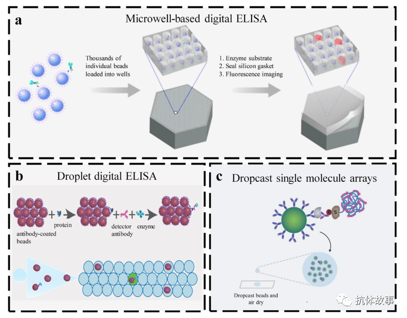
Figure 6. Three representative types of digital ELISA
Digital ELISA combines ELISA with digital counting for absolute quantification of target proteins. The first reported digital ELISA used MBs modified with specific antibodies to capture targets, followed by an enzyme-labeled immunocomplex capable of catalyzing substrates to produce fluorescent products. Finally, these MBs are dispersed into a series of ascending-sized wells, and single protein molecules are detected using fluorescence imaging technology. This method can detect about 10 to 20 enzyme-labeled complexes in a 100 μL sample, with a detection limit of 14 fg/mL.
Digital ELISA has been used to detect various low-abundance proteins, including tau associated with Alzheimer’s disease, HIV p24 antigen, cytokines, and influenza A nucleoprotein. The high sensitivity of digital ELISA provides new insights into disease development. However, digital enzyme-linked immunosorbent assays are still a type of sandwich enzyme-linked immunosorbent assay, requiring targets that can bind to a pair of antibodies. Therefore, digital ELISA for detecting small molecules (such as haptens) has not yet been reported. Additionally, non-specific adsorption of beads, enzyme labels, surfaces, and other reagents can easily generate background signals, posing significant challenges for researchers.
7. Conclusion and Future Prospects
ELISA is a fundamental tool for diagnosing IVD, widely applied in preliminary screening, therapeutic monitoring, and prognostic assessment of various diseases. However, its limited sensitivity, expensive instruments, and the need for trained professionals restrict its widespread application. This article discusses various new ELISA-derived technologies and compares their detection performance in terms of sensitivity, specificity, dynamic range, multi-component detection capability, reproducibility, practicality, and cost. All these technologies address one or more issues in protein detection. Although current POC devices such as lateral flow immunoassays have achieved on-site immunoassays, their sensitivity and multiplexing capabilities still do not meet user demands. Therefore, developing sensitive, multiplexed, and high-throughput POC immunoassay methods remains a major development direction for resource-limited areas. In addition to low-cost, rapid, and semi-quantitative analyses that do not require expensive instruments, some new ELISA-derived technologies also exhibit high sensitivity, making them particularly suitable for POC testing. However, these methods still require manual operations and lack multiplex analysis capabilities. Therefore, combining these ELISA with microfluidic technology can automate ELISA and enhance its multiplex detection capabilities. The second development direction is the optimization and miniaturization of digital ELISA. Currently, the sensitivity of digital ELISA (SIMOA) can reach single-molecule levels and can achieve multiplex detection of six indicators, making it quite powerful for diagnosing many clinical diseases. However, three shortcomings still need to be addressed. First, Poisson noise remains a significant concern when detecting low-concentration target molecules in digital analysis. Second, non-specific binding between samples, reagents, and surfaces may lead to inaccurate detection results. To address this, some researchers have developed surface passivation methods and reagents to reduce non-specific binding. Meanwhile, the mechanisms of non-specific binding require further investigation. Finally, due to the complexity of microtiter chips, nanoparticles, or microfluidic chips, the current costs of digital ELISA remain high. Therefore, it is necessary to develop a simple, low-cost digital ELISA method in the future. Finally, combining ELISA technology with emerging technologies such as new sensing technologies and new materials will promote its further development and expand its application scope. In summary, the development of sensitive ELISA biomolecular detection technology for IVD is a multidisciplinary research effort that relies on the integrated development of signal amplification technologies, advanced materials, and miniaturized devices.
【Original Source】
Peng P, Liu C, Li Z, et al. Emerging ELISA derived technologies for invitro diagnostics[J]. TrAC Trends in Analytical Chemistry, 2022, 152:116605-.
Original link: https://www.sciencedirect.com/science/article/pii/S0165993622000887
Instructor: Wang Zhanhui
Share your antibody stories and research progress on antibody-based analytical methods, welcome submissions!
Long press the image below to recognize the QR code and easily follow!