7T Ultra-High Field MRI Analysis of Human Brain β-Amyloid and Iron Deposition Characteristics

7T Ultra-High Field MRI Analysis of Human Brain β-Amyloid and Iron Deposition Characteristics

[Neuroscience Cutting-Edge Technology Training Series] See details at the end

[Content Promotion, Lectures/Reports/Courses and Other Academic Collaborations] Please contact WeChat: Wang_Sizhen

Source | Peking University Brain Science

Editor | Wang Sizhen

Alzheimer’s disease‘s core pathological feature is the formation of amyloid plaques caused by Aβ aggregation, while recent years have found that abnormalities in iron homeostasis are closely related to the pathogenesis of AD, a key factor in disease progression. From the perspective of magnetic resonance imaging physical processes, iron deposition signals exhibit paramagnetism, while signals from myelin, Aβ and other proteins primarily show diamagnetism.Traditional quantitative magnetic susceptibility imaging (QSM) technology mainly reflects paramagnetic effects related to changes in iron content, failing to effectively distinguish between the two coexisting signal sources of paramagnetism and diamagnetism within a voxel (voxel), and cannot quantitatively assess the levels of Aβ aggregation and iron deposition in brain tissue.
Recently, a collaboration among Zhejiang University School of Physics He Hongjian, Shanghai Jiao Tong University School of Biomedical Engineering Wei Hongjiang, Peking University IDG McGovern Institute for Brain Science, School of Psychological and Cognitive Sciences, Peking University-Tsinghua Life Science Joint Center Wang Zheng team has published an academic paper titled“Distinct regional vulnerability to Aβ and iron accumulation in postmortem AD brains” online in the internationally recognized professional journal“Alzheimer’s & Dementia”.This research integrates 7T ultra-high field quantitative magnetic susceptibility imaging, immunohistochemistry, and transcriptomics technologies, and quantitatively compares the spatial distribution patterns of βamyloid protein (Aβ) aggregation and iron deposition in human brain specimens from healthy volunteers and Alzheimer’s (AD) patients from multiple angles including gene expression, signaling pathways, and tissue pathology, discovering differences in susceptibility between the two in different brain regions, providing new insights into understanding the pathological mechanisms of AD, and opening new avenues for the application of 7T magnetic resonance imaging technology in the early diagnosis of AD.(Further reading: Wang Zheng‘s research progress, see “Logical Neuroscience” report (click to read):Adv Sci | Wang Zheng/ Wang Yingwei/ Wang Meiyun’s team reveals the brain network and transcriptional characteristics induced by electrical stimulation of the thalamic plate in macaques.Neurosci Bull | Wang Zheng/ Wang Meiyun’s team reveals the correlation between quantitative expression of individual implicit disease components and dimensions of psychopathology and treatment response.NeuroImage | Wang Zheng/ Zhao Min/ Li Biao’s team reveals the metabolic and functional mechanisms of cognitive decision-making abnormalities in addicted patients using PET/MRI imaging and behavioral computational modeling.Cell Reports | Peking University Wang Zheng’s team reveals that non-coding genes regulate the brain’s resting state functional network, which is highly associated with human mental illness risk genes.
7T Ultra-High Field MRI Analysis of Human Brain β-Amyloid and Iron Deposition Characteristics
In response to these multiple technical challenges, the research team first innovatively proposed a sub-voxel quantitative magnetic susceptibility imaging method (APART-QSM), constructing an accurate imaging physical model to distinguish between the paramagnetic and diamagnetic signals of brain tissue, used to quantify the levels of iron deposition and Aβ aggregation within the voxel, and subsequently collaborated deeply with the National Health and Disease Human Brain Tissue Resource Bank of Zhejiang University, conducting 7T ultra-high field magnetic resonance imaging and immunohistochemical analysis on post-mortem human brain samples donated by AD patients and healthy volunteers. It is worth mentioning that post-mortem brain tissue specimens are an important experimental model for exploring the mysteries of the brain, Wang Zheng‘s laboratory has long focused on the application of neuroimaging and neuroregulation techniques in brain science and brain diseases, and has accumulated rich experience in non-human primate post-mortem brain models.(Neuron, 2013; Cereb Cortex, 2021; eLife, 2022; Nat Commun, 2023; Advanced Science, 2024).

In this study, the research team conducted Aβ immunohistochemical staining and Perls/DAB staining on key pathological brain regions including the frontal gyrus, inferior parietal lobule, posterior hippocampus, parahippocampal gyrus, and entorhinal cortex of post-mortem human brain tissue, obtaining quantitative information on microscopic pathology, and through joint statistical analysis with macroscopic magnetic susceptibility, revealing the spatial distribution patterns of Aβ aggregation and brain iron levels. Finally, further integrating the human brain transcriptome map released by the Allen Institute for Brain Science (Nature, 2012), it deeply interprets the genetic characteristics behind Aβ and brain iron deposition, with Figure 1 illustrating the logical thinking and methodological flow of the overall experimental design.
7T Ultra-High Field MRI Analysis of Human Brain β-Amyloid and Iron Deposition Characteristics
Figure 1. Human Brain Sub-Voxel Quantitative Magnetic Susceptibility Imaging, Immunohistochemistry, and Transcriptomics Integrated Cross-Scale Analysis Route Map.
The research team analyzed the sub-voxel signal model and found that the region distribution representing AB protein aggregation signals|Xdia| has significant spatial consistency with the region distribution of iron deposition signalsXpara, reflecting the spatial co-localization of Aβ and iron deposition in the brain of AD patients. At the same time, specific distribution patterns of Aβ and iron were also discovered in the brain, which are difficult to achieve with traditional magnetic resonance imaging technology: the diamagnetic changes in the medial prefrontal cortex are greater than the paramagnetic changes, indicating that the accumulation level of Aβ in these regions may be relatively higher; conversely, in the central frontal gyrus, medial frontal gyrus, and temporal lobe cortex, paramagnetic changes are greater than diamagnetic differences, corresponding to more significant pathological effects of iron deposition.Figure 2.
7T Ultra-High Field MRI Analysis of Human Brain β-Amyloid and Iron Deposition Characteristics
Figure 2. Spatial Distribution Map of Differences in Aβ and Iron Deposition between AD Patients and Healthy Controls in the Brain Cortex. (a) and (b) are the results of two-sample t-tests between the two groups, representing the distribution patterns of Aβ and iron deposition in the cortex of AD brains.(c) Scatter plot of regional diamagnetic differences and paramagnetic differences shows significant positive correlation, with each data point representing one of the 168 cortical regions, blue and red markers represent regions with significant differences in Aβ and iron deposition.
Intracerebral Aβ protein and iron deposition are believed to be closely related to genetic factors. Therefore, the research team combined the above quantitative magnetic susceptibility brain region distribution difference maps with Allen Human Brain Transcriptome Map data to establish a partial least squares regression model for imaging-transcriptomics analysis. The results found that the accumulation of Aβ proteins in regions such as the prefrontal cortex, inferior parietal, and occipital lobes was significantly correlated with the expression of 2408 genes, most of which were associated with processes such as cell cycle, cellular stress response, and protein metabolism regulation; while the spatial distribution patterns of iron deposition were significantly correlated with the expression profiles of 2886 genes, in addition to cell cycle and cellular stress response, also associated with processes such as cell death, revealing the similarities and differences in their potential contributions to the pathogenesis of AD.(Figure 3)
7T Ultra-High Field MRI Analysis of Human Brain β-Amyloid and Iron Deposition Characteristics
Figure 3. Gene Enrichment Analysis Related to Iron Deposition and Aβ Aggregation in Alzheimer’s Disease. (a) Pathways related to iron deposition. (b) Pathways related to Aβ aggregation. These pathways are plotted in semantic space, with more similar items clustering together. Only non-redundant items that are significant at g:SCS correction P < 0.001 are shown. Larger and deeper circles indicate greater significance; (c) Overlap of pathways between the two.
Article Conclusion and Discussion, Inspiration and Outlook

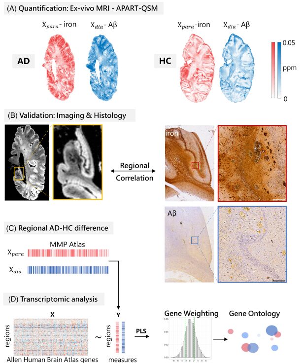
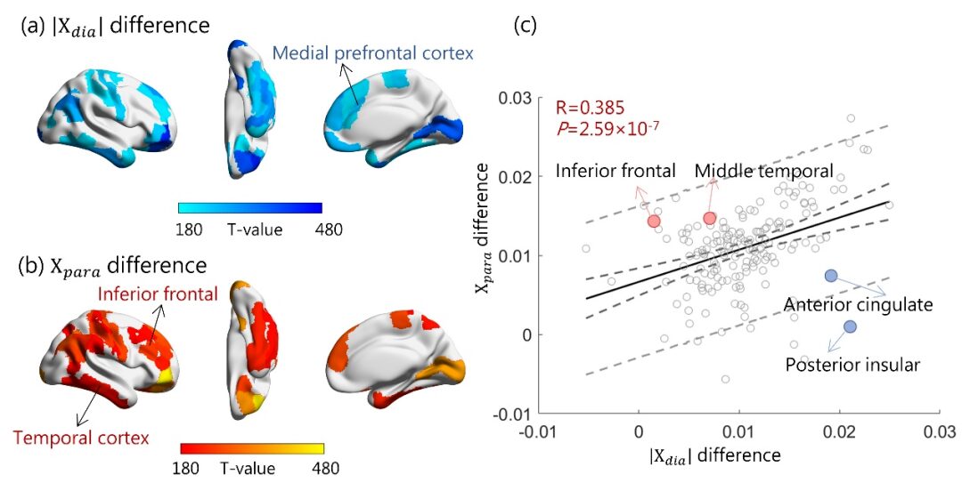
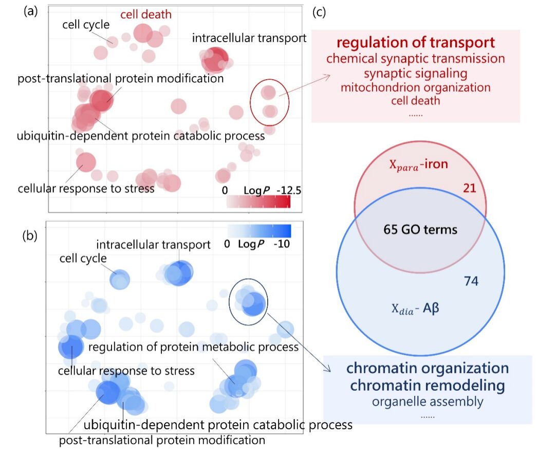
This study simultaneously excavates the rich information contained in post-mortem human brain specimens from both macro imaging and micro pathology scales and incorporates transcriptomic analysis, analyzing the gene expression characteristics behind brain Aβ protein and iron deposition, which is of great significance for deepening the pathological research of AD. It is worth emphasizing that the innovative 7T ultra-high field magnetic resonance imaging technology can analyze the changes in diamagnetic and paramagnetic susceptibility in quantitative magnetic susceptibility signals, and shows a close correlation with the pathological progression of AD, suggesting the potential clinical application value of quantitatively assessing the two magnetic susceptibility characteristics, which may provide new imaging indicators for the early clinical diagnosis of AD, offering better cost-effectiveness compared to existing PET molecular imaging diagnosis, benefiting a wider patient population.

Original linkSee:https://alz-journals.onlinelibrary.wiley.com/doi/10.1002/alz.14188
Zhejiang University School of Biomedical Engineering and Instrument Science PhD student Yao Junye is the first author of this article. Peking University Wang Zheng, Zhejiang University He Hongjian, Shanghai Jiao Tong University Wei Hongjiang are the corresponding authors of this article. Shanghai Jiao Tong University PhD student Li Zhenghao and Stanford University postdoctoral fellow Zhou Zihan made significant contributions to this work, and Zhejiang University Bao Aimin professor and his team provided valuable guidance and suggestions for this research. This research was jointly funded by the Ministry of Science and Technology’s Science and Technology Innovation 2030-“Brain Science and Brain-Like Research”, the National Natural Science Foundation, and Peking University-Tsinghua Life Science Joint Center and other projects.

Reprint Notice:Non“Logical Neuroscience” team original manuscripts and (or) invited manuscripts, this content is authorized for reprint, copyright belongs to the original author and (or) original unit..

7T Ultra-High Field MRI Analysis of Human Brain β-Amyloid and Iron Deposition Characteristics
[Neuroscience Cutting-Edge Technology Training Series]

[Optogenetics and Genetic Encoding Calcium Probes and Neurotransmitter Probe Working Principles and Applications] and [In Vivo Imaging Techniques in Neuroscience Research Basics and Applications] (Issue 9), time March 22-24, 2025 (Saturday to Monday); location Nanjing.

[Patch-Clamp Recording System Operation and Application] (Issue 7). Time To be determined; location Nantong.

[Neuroscience Cutting-Edge Technology Integration: When In Vivo Electrophysiology Meets Optogenetics] (Issue 2), time To be determined (Saturday to Monday); location Nanjing.

[National Frontier Tissue Transparency and Three-Dimensional Imaging Theory and Skills Training Course] (Issue 2), time To be determined; location Wuhan.

[Disease Research and Animal Behavior Series]

[Latest Research Progress and Hotspots of Alzheimer’s Disease and Experimental Design Ideas and Data Analysis of Animal Behavior] (Issue 2). Time March 23, 2025 (Sunday), location Online.

“Theoretical Knowledge, Operational Skills, Scientific Research Thinking”
Registration method:15108204441 (phone),Wang_Sizhen (WeChat)
(Click title to read the full text)

“Logical Neuroscience” WeChat Group: Literature Study

Scan to add WeChat, and note: LogicalLiteratureNameUnitResearch FieldDegree/Title
(Note: If not noted in the required format, the friend request will not be approved)
7T Ultra-High Field MRI Analysis of Human Brain β-Amyloid and Iron Deposition Characteristics
References
1. Wang Z et al., The relationship of anatomical and functional connectivity to resting state connectivity in primate somatosensory cortex, 2013, Neuron
2. Lv QM et al., Normative Analysis of Individual Brain Differences Based on a Population MRI-Based Atlas of Cynomolgus Macaques, 2021, Cerebral Cortex.
3. Yan MC et al., Mapping brain-wide excitatory projectome of primate prefrontal cortex at submicron resolution and comparison with diffusion tractography, 2022, eLife.
4. Bo TT et al., Brain-wide and cell-specific transcriptomic insights into MRI-derived cortical morphology in macaque monkeys, 2023, Nature Communications.
5. Zhang Z et al., State-specific regulation of electrical stimulation in the intralaminar thalamus of macaque monkeys: network and transcriptional insights into arousal, 2024, Advanced Science.
6. Hawrylycz MJ, et al., An anatomically comprehensive atlas of the adult human brain transcriptome, 2012, Nature.
Editor | Wang Sizhen
The End of This Article

Leave a Comment