

 Introduction: Continuing the discussion on instrument-assisted diagnosis, today we will talk about the cast type that is second only to the transparent cast type – the granular cast type. The characteristics under the microscope can be understood as the presence of >1/3 granules (whether fine or coarse granules) in the transparent cast type. Let’s take a look at this small case together.
  PART01: Patient: Male, 25 years old, with traumatic subarachnoid hemorrhage, urine routine examination results as follows:★ Looking at the results below, the regular record indicates that only pathological casts are visible (all types other than transparent casts are classified as pathological casts – unclassified casts). Since image-based urine sediment analyzers have increased the detection sensitivity for casts, their specificity may be relatively poor, so careful observation is needed to determine if they are truly pathological casts.
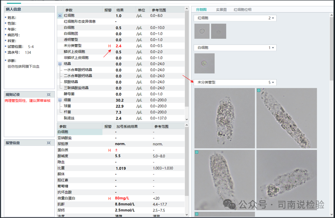 ★ Check the unclassified cast area:
 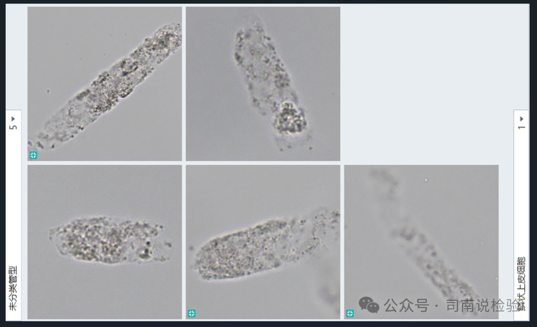 ★ Granular cast components are visible, with several magnified:
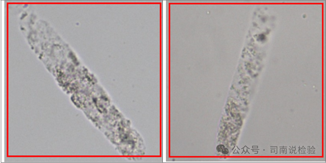 ★ For abnormal results (including those triggering rules and those considered potentially abnormal), centrifugation and microscopy are required:
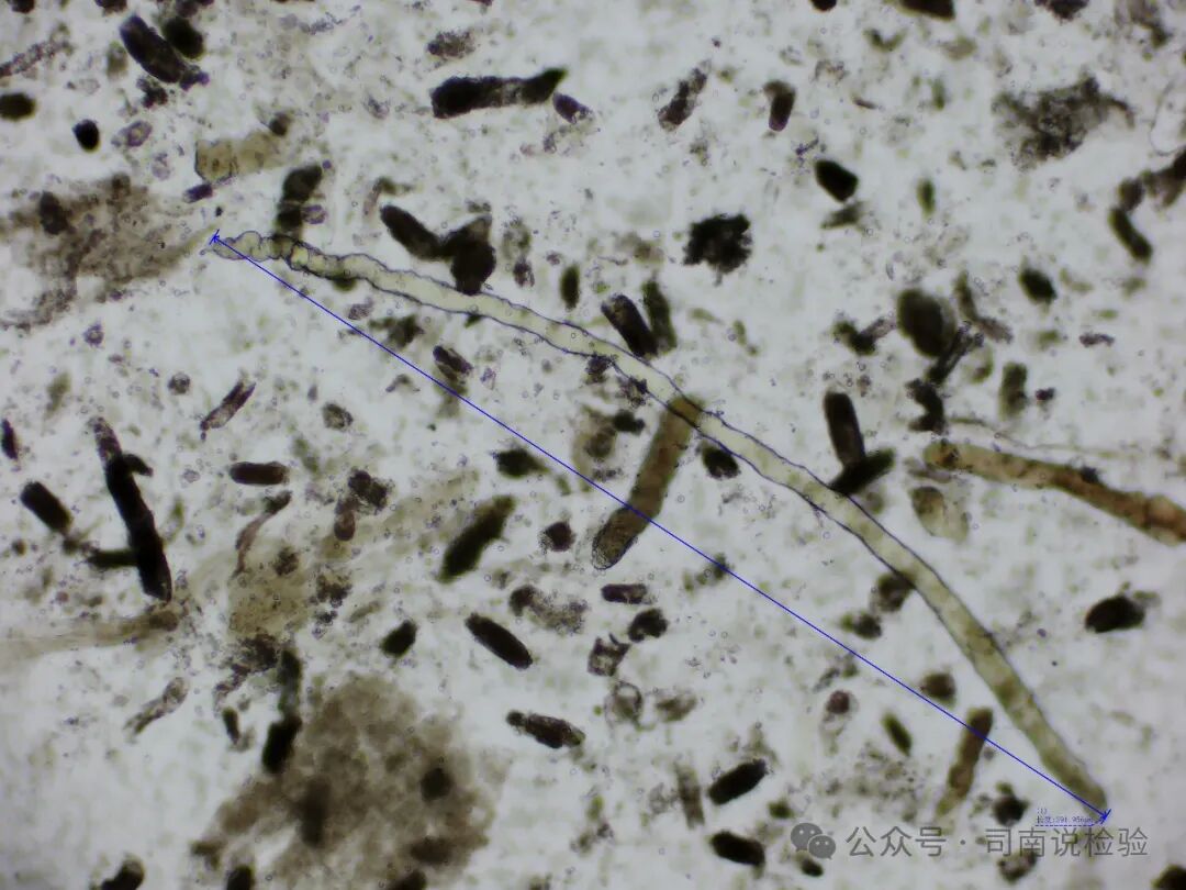
 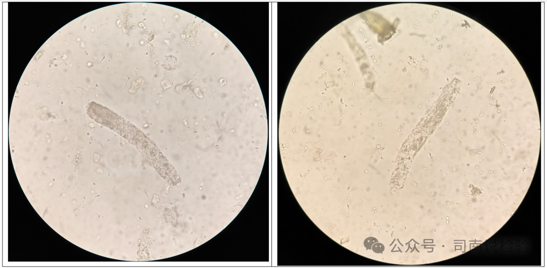 ★ Granular casts are visible under the microscope, and the instrument’s capture is accurate. Today’s content is a bit short, so here’s a question: what type of casts are these below?
 ★ Knowledge Expansion: Granular Casts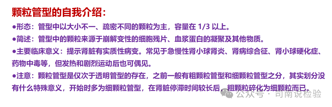
 Si Nan says: Casts can be understood morphologically as transparent casts + issues; granular casts are simply transparent casts + internal granules >1/3. Granular casts are clearly defined in textbooks, but in practical work, they can sometimes be difficult to distinguish from fatty casts (unstained), blood casts (darker in color), and muddy brown casts (with very deep granules). These require careful observation and thought. Additionally, the distinction between fine granular casts and coarse granular casts is not very significant; they can be classified under the same meaning. Lastly, it is still true that in clinical testing work, details determine success or failure; testing should seek truth, and verification should uphold integrity! Note: Due to the author’s limited knowledge and experience, this article mainly organizes past materials and experiences from practical applications, intended for communication, learning, and sharing purposes only. The content should not be used as a clinical diagnostic basis. If there are any errors, please contact the author privately. This is for non-commercial use only, intended for reference and communication among professionals. If there are any infringements, please contact the author for deletion. Thank you! References:1. Atlas of Clinical Body Fluids and Excretions Morphological Examination, Wang Qian, Zheng Lei, Sun Dehua (Editors)2. Color Atlas of Urine Sediment, Zhang Yunhu3. New Interpretation and Case Analysis of Urinary Formed Elements Atlas, Yan Lizhi4. Comprehensive Analysis of Urine and Urine Sediment Atlas, Cao Yan5. Expert Consensus on the Names and Result Reports of Urinary Formed Elements by the Hematology and Body Fluids Group of the Chinese Medical Association Laboratory Medicine Branch. Journal of Chinese Laboratory Medicine, 2021, 44(7): 574-586.6. Mindray Instrument Manual7. Public Account: Zhang Shimin’s Academic Classroom

In fact, good things and suffering are just two sides of the same coin; one is the back of the hand, and the other is the palm. If one can truly let go of good things, then suffering can also be let go of. Therefore, suffering is also a good test. If a person cannot let go when encountering troubles, pain, or adversity, it is impossible for them to let go when encountering good things.
————– Nan Huai Jin