 ▲ Design and Function of Biohybrid Microbots for Active Pneumonia TreatmentThe full text contains6767words, reading takes about15minutes
-
Pneumonia, as a global public health challenge, still lacks efficient and precise treatment methods. Yu Yingfeng from Southern Medical University, Peng Fei from Sun Yat-sen University, and Shen Lihan from Dongguan People’s Hospital have combined magnetotactic bacteria with biomimetic cell membrane nanoparticles for the first time, constructing a magnetically controlled biohybrid microbot that achieves multiple functions including neutralization of inflammatory factors, viral blockage, and active drug delivery. Under the influence of a magnetic field in the lungs, this microbot demonstrates excellent retention and therapeutic effects in the lungs, opening up a new path for precise treatment of complex respiratory infections.
Written/Edited by|shanjie@ahmu
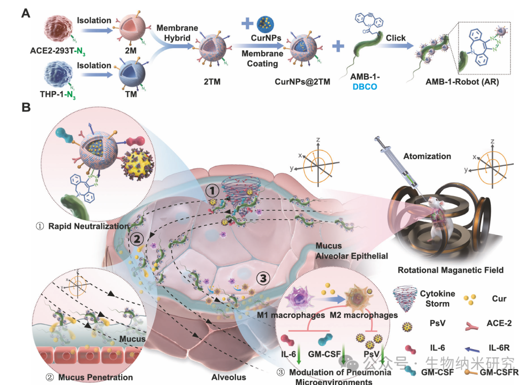
In the past decade, significant progress has been made in the design of synthetic micro/nano robots with various functions, showing great potential in biomedical applications. However, there remains a significant gap in achieving efficient treatment from in vitro to in vivo. In this study, Yu Yingfeng from Southern Medical University, Peng Fei from Sun Yat-sen University, and Shen Lihan from Dongguan People’s Hospital utilized “click chemistry” to modify curcumin-loaded hybrid cell membrane nanoparticles onto the surface of magnetotactic bacteria AMB-1 (Magnetospirillum magneticum AMB-1), and combined it with magnetic driving to construct the biohybrid microbot CurNPs@2TM AMB-1 for active and efficient pneumonia treatment in vivo. Under the influence of an external rotating magnetic field, this hybrid robot can achieve controllable, wireless-driven movement and efficiently eliminate inflammatory factors and SARS-CoV-2 pseudovirus, thereby inhibiting viral invasion and reducing lung damage caused by inflammation. Due to its active movement characteristics, this robot can significantly increase and prolong the accumulation of curcumin in the lungs, effectively alleviating pneumonia and regulating the pneumonia microenvironment. This hybrid microbot system, which possesses magnetic driving, active neutralization of inflammatory factors, and viral interception capabilities, provides a highly promising new therapeutic platform for pneumonia treatment. The related research was published in Biomimetic magnetobacterial microrobots for active pneumonia therapy in Nature Communications.1
Preparation of AR
Construction of biomimetic N3 labeled cell membrane coated nanoparticles
First, biomimetic N3 labeled cell membrane coated nanoparticles (CurNPs@2TM) were constructed. As shown in Fig. 2A, the researchers pre-treated ACE2-293T cells and THP1 cells with N3 functionalized Ac4GalNAz, and the N3 functional group was introduced through the sugar metabolism pathway. To verify the presence of the N3 group, fluorescent DBCO-Cy5 was added to the cells, and the reaction between DBCO and N3 was achieved using click chemistry, followed by imaging observation using fluorescence microscopy (Fig. 2B, C). Compared to untreated cells, the cells pre-treated with Ac4GalNAz exhibited significant red fluorescence signals. Further validation using flow cytometry confirmed the successful introduction of the N3 group on the surface of THP1 cells and ACE2-293T cells (Fig. 2D–I and Supplementary Fig. 1).
After completing the N3 labeling, the ACE2-293T cell membrane (2M) and THP1 cell membrane (TM) were extracted and fused to form the N3 functionalized 2M-TM hybrid cell membrane (2TM). Meanwhile, human serum albumin (HSA) and curcumin (Cur) were assembled through ultrasound to synthesize CurNPs, and then the 2TM was coated onto the surface of CurNPs using a repeated extrusion method, resulting in CurNPs@2TM. Further, 2M was labeled with DII, and TM was labeled with DID, and then the fluorescently labeled hybrid cell membrane was coated onto CurNPs through the extrusion method, and observed under an inverted fluorescence microscope. The results showed that the fluorescence of 2M and TM largely overlapped with the fluorescence of Cur, indicating that CurNPs@2TM was indeed composed of hybrid cell membranes (Supplementary Fig. 2).
As shown in Fig. 2J, transmission electron microscopy (TEM) observed a distinct core-shell structure, with a cell membrane thickness of about 25 nm. The particle size of CurNPs@2TM increased from 121 ± 3.05 nm to 161 ± 2.50 nm after coating, and the distribution was relatively narrow (Supplementary Fig. 3). Its Zeta potential decreased from −19.8 ± 0.40 mV to −29.0 ± 0.92 mV (Supplementary Fig. 4), indicating successful coating of negatively charged cell membranes. Western blot analysis further confirmed that CurNPs@2TM retained key membrane receptors, including ACE2 for neutralizing SARS-CoV-2, CD116 for neutralizing GM-CSF, and IL6ST for neutralizing IL-6 (Fig. 2K and Supplementary Fig. 25), indicating effective modification of CurNPs by the 2TM hybrid cell membrane.
Modification of magnetotactic bacteria and assembly of AR
The natural magnetotactic bacteria AMB-1 were cultured in enriched magnetospirillum growth medium (EMSGM). To achieve DBCO modification, DBCO-PEG4-NHS ester was added to AMB-1, and DBCO labeled AMB-1 was obtained through esterification reaction (Fig. 2L). Subsequently, the CurNPs@2TM with N3 groups were coupled with DBCO labeled AMB-1 through click chemistry, successfully preparing AR.
Scanning electron microscopy (SEM) was further used to verify the surface modification of AMB-1. As shown in Fig. 2M, the unmodified AMB-1 surface was smooth, while after the click reaction with CurNPs@2TM, multiple covalently bound nanoparticles were visible on the bacterial surface. Since curcumin itself has fluorescence, the obtained AR exhibited uniform and strong fluorescence signals, which remained clearly visible even after multiple washes (Fig. 2N), indicating that CurNPs@2TM had been firmly modified on the surface of AMB-1. The introduction of CurNPs@2TM also slightly decreased the Zeta potential of AR from −16.2 ± 0.55 mV to −18.5 ± 0.41 mV (Supplementary Fig. 4).
The click chemistry reaction conditions were mild, with minimal impact on cell viability. After 1 day of reaction, only a few bacteria deaths were observed (Supplementary Fig. 5). Further assessment of bacterial survival found that after 1 day of reaction, the colony counts of AMB-1 and AR were close, with an AR survival rate of about 95% (Supplementary Fig. 6). To study the survival time of AMB-1 after loading CurNPs@2TM, the growth curve of AR was plotted, and the results showed that its growth was only slightly affected, with no significant difference compared to the control, indicating that magnetotactic bacteria could survive normally after loading CurNPs@2TM (Supplementary Fig. 7).
Physicochemical properties of AR
Further studies on the physicochemical properties of AR were conducted. UV-visible absorption and fluorescence spectra showed that AR exhibited absorption and emission peaks at 432 nm and 544 nm, respectively, consistent with the spectral characteristics of CurNPs@2TM (Supplementary Fig. 8–9). The effective fluorescence intensity of curcumin-loaded AR is also significant for in vivo imaging, which can be used to monitor the accumulation of AR.
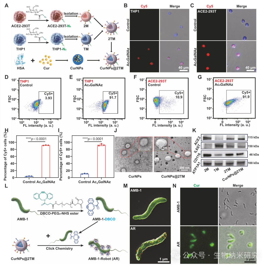
Figure 2 | Preparation and characterization of CurNPs@2TM and AR
2
Movement Behavior of AR
After successfully preparing AR, the researchers further studied the movement behavior of this biomimetic magnetic bacterial microbot in simulated lung fluid (SLF) under an external rotating magnetic field (RMF) generated by a self-made three-dimensional Helmholtz coil system. As shown in Fig. 3A, the applied RMF can convert rotational motion into translational propulsion. As a magnetotactic bacterium, AMB-1 has a diameter of about 0.5 μm and a length of about 3 μm, with intracellular magnetosomes averaging about 50 nm in diameter, which are arranged in a chain along the bacterial motion axis, consistent with previous reports (Fig. 3B), providing the premise for magnetic driving.
The researchers evaluated the magnetic properties of AMB-1 and AR using a vibrating sample magnetometer (VSM). The hysteresis loops indicated that both AMB-1 and AR exhibited superparamagnetism, with saturation magnetization (Ms) of 1.82 emu/g and 1.71 emu/g, respectively (Fig. 3C), suggesting that the superparamagnetic properties of AMB-1 were maintained after the introduction of CurNPs@2TM.
Subsequently, AMB-1 and AR were added to a confocal culture dish, and their movements were recorded using an inverted microscope, followed by motion trajectory analysis using ImageJ software. The results showed that in SLF, the forward speed of AMB-1 and AR was positively correlated with the rotation frequency of the 7 mT magnetic field, with speed gradually increasing until the frequency reached 24 Hz, accompanied by increased displacement (Fig. 3D). However, when the frequency exceeded 24 Hz, AMB-1 and AR could no longer synchronize with the rotating magnetic field, leading to a significant decrease in propulsion speed, confirming 24 Hz as their “loss of step frequency.” This desynchronization phenomenon may be due to the dominance of viscous resistance over magnetic torque at high frequencies.
Figures 3E–G show the representative movement trajectories of AMB-1 and AR at different input frequencies, indicating that both movements exhibit directionality and controllability. As shown in Fig. 3F, H, the mean square displacement (MSD) follows a parabolic fitting curve, further indicating that AMB-1 and AR achieved directional movement under magnetic field control. Compared to AMB-1, the forward speed of AR slightly decreased due to the introduction of CurNPs@2TM (Fig. 3D, Supplementary Fig. 10–11).
Meanwhile, by adjusting the RMF, the researchers were able to precisely guide AR along user-defined trajectories at the microscale. As shown in Fig. 3I, J and Supplementary Movie 1, AR successfully navigated a “W” shaped path under real-time control. These results indicate that AR possesses highly controllable and efficient movement capabilities, which is a key prerequisite for its future application in pneumonia magnetic microbot therapy.
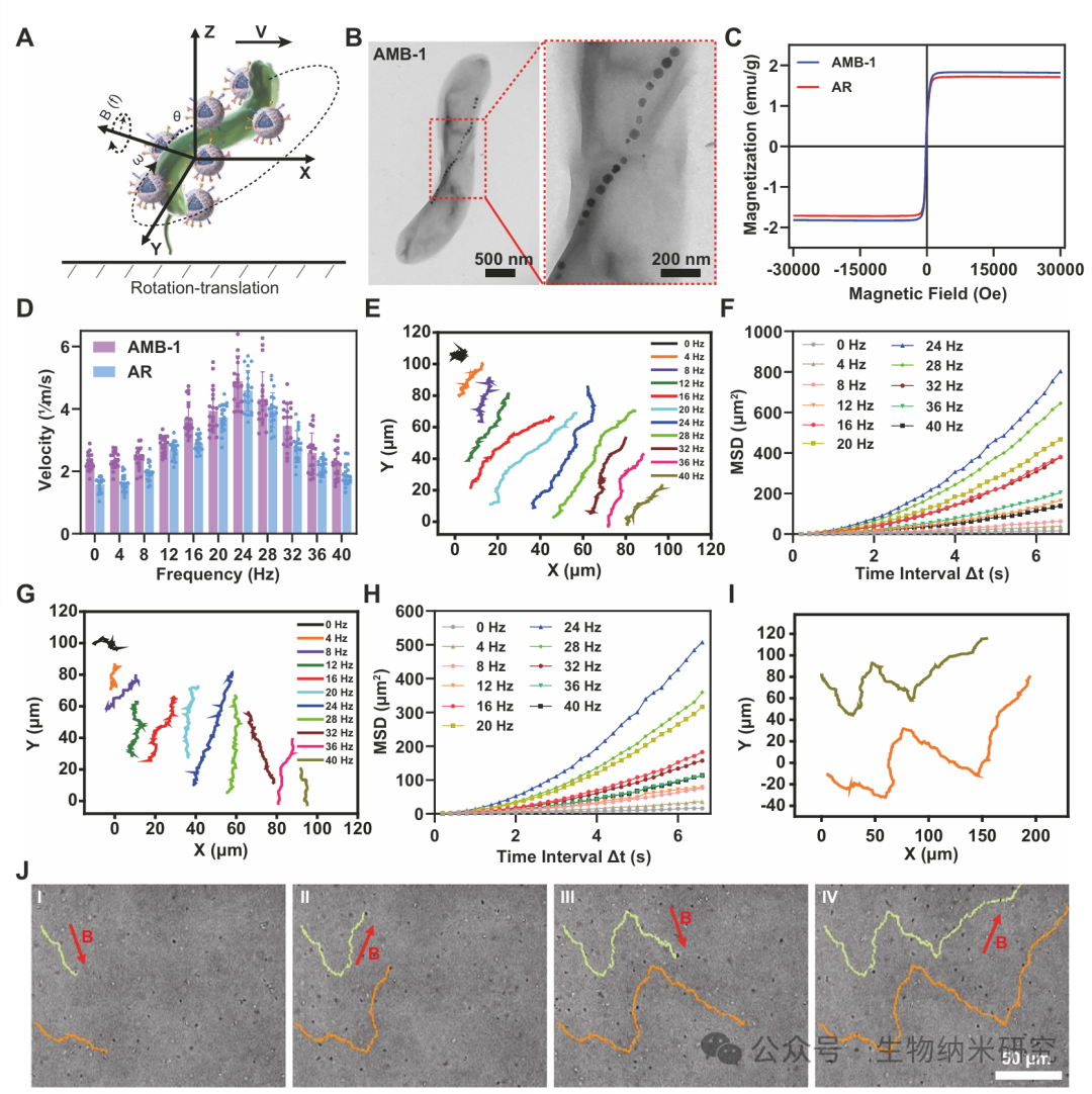
Figure 3 | Magnetic Driving and Motion Control of AR
3
AR’s In Vivo Lung Retention
Before evaluating its pneumonia treatment effect, the researchers first studied the lung retention of AR in C57 mice. The mice were randomly grouped and underwent intratracheal (i.t.) inhalation of different samples. For the magnetic driving (+M) group, the mice were placed at the center of a self-made three-axis Helmholtz coil after 1 hour of i.t. inhalation and received 30 minutes of magnetic field treatment (Fig. 4A). Benefiting from the active movement in the alveoli under RMF driving, AR was believed to penetrate the lung mucus and promote its retention in the lungs (Fig. 4B).
To study how magnetic field-driven movement enhances AR’s penetration of lung mucus, the researchers labeled AR with Cy5 fluorescence and administered it via i.t. inhalation into the mice’s lungs. After magnetic field treatment, lung tissue sections were observed, and the results showed that under RMF, AR could effectively penetrate lung mucus and reach the lung epithelium (Supplementary Fig. 12). Based on this, the researchers speculated that the translational helical motion induced by the three-axis Helmholtz coil helps promote AR’s penetration of lung mucus, enabling subsequent drug delivery to lung epithelial cells.
Subsequently, the researchers synthesized ICGNPs@2TM-AMB-1 by replacing curcumin with indocyanine green (ICG) with an excitation wavelength in the near-infrared region, followed by hybrid cell membrane coating and AMB-1 coupling. Utilizing the fluorescence characteristics of ICG, the lung distribution of mice after i.t. administration was evaluated at different time points using an in vivo imaging system (IVIS). As shown in Fig. 4C, compared to the ICGNPs@2TM group alone, the mice receiving ICGNPs@2TM-AMB-1 combined with magnetic field treatment (ICGNPs@2TM-AMB-1+M) exhibited stronger fluorescence signals and retained for at least 72 hours. In contrast, the signal of the ICGNPs@2TM group rapidly diminished within 24 hours, becoming nearly undetectable by 48 hours, highlighting the critical role of active movement in enhancing lung distribution and retention. Further semi-quantitative analysis indicated that the fluorescence intensity of the ICGNPs@2TM-AMB-1+M group was the highest, more than twice that of the group without magnetic field treatment (Fig. 4D). Normalized fluorescence data further verified that the clearance rate of ICGNPs@2TM-AMB-1+M was slower.
After 72 hours of administration, the researchers euthanized the mice and collected organs for ex vivo imaging. The results showed that the fluorescence of ICGNPs@2TM in lung tissue was almost undetectable, while the ICGNPs@2TM-AMB-1+M group exhibited the best retention effect in the lungs (Fig. 4E), benefiting from the active movement performance driven by RMF. Semi-quantitative fluorescence analysis further indicated that the retention amount of ICGNPs@2TM-AMB-1+M in lung tissue was about 3 times that of the group without magnetic driving (Fig. 4F), suggesting that magnetic driving movement can enhance its penetration of lung mucus and promote the accumulation of magnetic bacterial microbots.
Additionally, the researchers evaluated the release and retention of curcumin from AR. After 72 hours of i.t. inhalation, lung tissue sections were collected for observation. As shown in Fig. 4G, compared to CurNPs@2TM and AR without RMF application, the lung tissue of mice treated with AR under RMF control showed stronger fluorescence signals, further proving its more efficient drug release and retention capabilities.
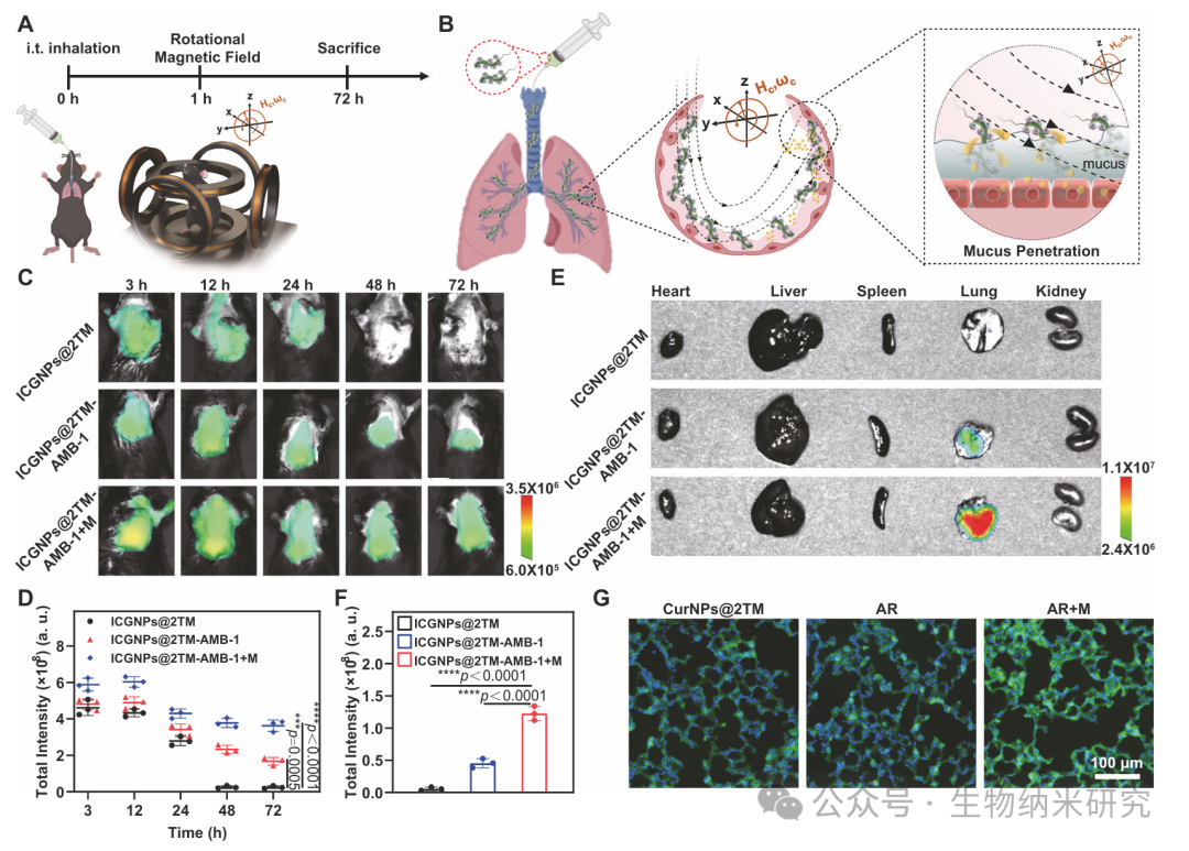
Figure 4 | Lung Distribution and Retention of AR
4
In Vitro PsV Inhibition and Inflammatory Factor Neutralization by AR
After confirming that AR could retain in the lungs under magnetic field influence, the researchers further evaluated its neutralization effect on viruses and inflammatory factors. Due to the coating of hybrid cell membranes, AR inherited abundant receptors from ACE2-293T cells and THP1 cells, including ACE2 for SARS-CoV-2 and cytokine receptors for IL-6 and GM-CSF, enabling it to neutralize viruses and inflammatory factors by competing for binding with host cells, thereby intervening in the occurrence and development of pneumonia (Fig. 5A, B).
First, the researchers assessed the biocompatibility of AR using the Cell Counting Kit-8 (CCK8) assay. AR with AMB-1 concentration of 10^8 cfu/mL and curcumin concentration of 10 μg/mL was co-cultured with ACE2-293T cells, and no significant cytotoxicity was observed (Fig. 5C), with cell viability greater than 90%, indicating that AR has excellent biocompatibility.
Subsequently, SARS-CoV-2 pseudovirus (PsV) with spike protein was synthesized and co-cultured with ACE2-293T cells in the presence of AMB-1, CurNPs@2TM, or AR. The results showed that cells co-treated with PBS or AMB-1 could be infected by PsV (Fig. 5D), due to the lack of ACE2 receptors. However, AR, due to the hybrid cell membrane coating and enrichment of ACE2, could compete with ACE2-293T cells for binding to PsV, effectively inhibiting infection, with inhibition effects comparable to CurNPs@2TM. This suggests that AR prepared by click chemistry coupling did not weaken its PsV neutralization ability.
The researchers further investigated the inflammatory factor neutralization effect of AR. AR was co-cultured with PBS containing known initial concentrations of IL-6 (2 μg/mL) and GM-CSF (0.5 μg/mL) for 30 minutes, and the residual factor concentrations in the supernatant were detected after centrifugation to remove AR. The results showed that both CurNPs@2TM and AR could efficiently neutralize IL-6 and GM-CSF (Fig. 5E, F), indicating that AR has the potential to alleviate the inflammatory storm. Although this system mainly targets IL-6 and GM-CSF, due to the broad-spectrum cytokine receptors expressed on the surface of AR, it also has application potential in neutralizing more pro-inflammatory factors.
In Vivo PsV Inhibition and Inflammatory Factor Neutralization by AR
After obtaining positive results from in vitro experiments, the researchers further validated in vivo. This study used the HACE2 mouse model, which overexpresses ACE2. The mice were randomly divided into six groups after pre-inhaling PsV via a micro-spray inhaler: PBS group, AMB-1 group, AMB-1+M group, CurNPs@2TM group, AR group, and AR+M group. After 0.5 h of PsV administration, AMB-1, CurNPs@2TM, or AR were administered via i.t. inhalation, with the AMB-1+M group and AR+M group placed at the center of the Helmholtz coil for 30 minutes of magnetic driving after 1 hour of inhalation (Fig. 5G).
Real-time in vivo bioluminescence imaging was used to monitor PsV infection, with higher fluorescence intensity indicating more severe infection. The results showed that the AR+M group had the weakest luminescence signal (Fig. 5H, I), indicating its strongest PsV inhibition ability. Compared to the AR group, the total fluorescence signal of the AR+M group was significantly reduced, decreasing by about 10 times compared to the AR group.
Subsequently, histopathological analysis of lung tissues from the mice was performed. The results showed that PsV infection could induce pneumonia and cause significant morphological damage (Fig. 5J). Notably, compared to the AR group, the AR+M group significantly alleviated pneumonia symptoms. Further scoring of lung tissue sections revealed that AR treatment could alleviate PsV-induced lung damage to some extent, while the AR combined with magnetic driving (AR+M) exhibited significantly enhanced therapeutic effects, with the lowest lung damage score (Fig. 5K).
Additionally, the researchers collected bronchoalveolar lavage fluid (BALF) for cytokine detection. The results showed that IL-6 and GM-CSF levels significantly increased after PsV exposure, while AR+M treatment significantly reduced the levels of IL-6 and GM-CSF in BALF (Fig. 5L, M), proving its broad-spectrum neutralization potential for inflammatory factors in vivo. The researchers speculated that this effect arises from the enhanced contact of AR with inflammatory factors due to its magnetic driving movement in the alveoli, thereby improving neutralization efficiency. Meanwhile, the active movement of AR also promotes its penetration through the lung mucus layer, reaching lung epithelial cells to release curcumin, further enhancing the retention of the drug in the lungs. Under the action of curcumin, the release of inflammatory factors in the alveoli can also be further suppressed.
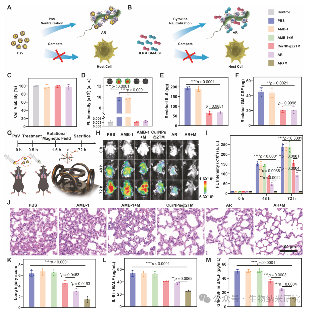
Figure 5 | In Vitro and In Vivo PsV Inhibition and Inflammatory Factor Neutralization by AR
5
AR Inhibits Acute Pneumonia
After confirming that AR+M possesses in vivo cytokine neutralization effects, the researchers further evaluated its ability to treat acute pneumonia. Acute pneumonia is an acute lung infection caused by pathogens such as bacteria or viruses, often accompanied by symptoms such as fever, cough, and difficulty breathing, requiring timely intervention. To establish an acute pneumonia model, the researchers treated C57 mice with intratracheal (i.t.) inhalation of lipopolysaccharide (LPS, 10 mg/mL). The LPS-pretreated mice were randomly divided into six groups: PBS group, AMB-1 group, AMB-1+M group, CurNPs@2TM group, AR group, and AR+M group. Subsequently, after 3 hours of LPS treatment, AMB-1, CurNPs@2TM, or AR were administered via i.t. inhalation. The AMB-1+M group and AR+M group were placed at the center of the Helmholtz coil for 30 minutes of magnetic driving after 1 hour of administration (Fig. 6A).
Curcumin has been shown to have ROS scavenging and anti-inflammatory effects and is widely used in the treatment of various inflammatory diseases. Therefore, the researchers assessed ROS production in acute pneumonia mice. First, 24 hours after LPS inhalation, the ROS-sensitive probe L-012 (25 mg/kg) was injected intravenously to evaluate lung oxidative stress, and inflammation was recorded using IVIS imaging. As shown in Fig. 6B, the PBS group lung tissues exhibited strong bioluminescence signals, while the AR+M group showed extremely weak signals, significantly lower than the AR group. This is because, under the magnetic field influence, AR enhanced the retention of curcumin in lung tissues, promoting ROS clearance. Semi-quantitative analysis of lung tissue luminescence intensity showed that the AR+M group reduced by about 12 times compared to the PBS group and about 6 times compared to the AR group, further indicating that AR+M can significantly alleviate LPS-induced acute pneumonia (Fig. 6C).
After 24 hours of LPS treatment, the mice were euthanized for histological analysis. The PBS group mice exhibited significant thickening of alveolar walls, disappearance of alveolar cavities, vascular dilation and congestion, and massive infiltration of inflammatory cells, indicating severe lung damage (Fig. 6D). Notably, AR+M significantly inhibited lung tissue damage, further proving the significant therapeutic effect of active AR under magnetic driving. Based on the Mikawa method, histological scoring showed that the PBS, AMB-1, and AMB-1+M groups had higher scores, while the AR+M group had the lowest damage score (Fig. 6E), demonstrating significant therapeutic effects.
Further, the lung wet/dry weight ratio (W/D) was measured to assess the degree of pulmonary edema in acute pneumonia mice. The lung W/D ratio reflects the water content of lung tissues, indirectly reflecting the degree of inflammation and edema. Acute pneumonia leads to increased exudation and elevated W/D ratios. The results indicated that the AR+M group had the largest decrease in W/D ratio, suggesting the highest degree of pneumonia alleviation (Fig. 6F).
The researchers also measured the levels of inflammatory factors in BALF. The results showed that IL-6 and GM-CSF levels significantly increased in the PBS group, indicating severe inflammatory status. The AMB-1 group and AMB-1+M group were similar to the PBS group, indicating that AMB-1 had no therapeutic effect. However, AR+M significantly reduced the levels of IL-6 and GM-CSF in BALF, with a clearance rate exceeding 85%, further demonstrating its effectiveness in pneumonia treatment (Fig. 6G, H). The above results indicate that under magnetic field influence, AR can achieve rapid neutralization of inflammatory factors in the pneumonia microenvironment, and by enhancing the retention and release of curcumin in the lungs, further suppress the production of inflammatory factors, effectively promoting pneumonia recovery.
Macrophage Polarization and Biocompatibility Evaluation
Reprogramming and polarization of pulmonary macrophages play a key role in the occurrence, development, and severity of pneumonia. Therefore, regulating the activation and differentiation of lung macrophages is considered a potential therapeutic strategy. Reducing macrophage infiltration or polarizing them to the M2 phenotype can effectively alleviate pulmonary inflammation.
To study the immune regulatory role of AR+M in the anti-inflammatory process of acute pneumonia, the researchers performed immunostaining on lung tissue sections from mice. F4/80 (red) and CD11c (green) were used to mark pro-inflammatory M1 macrophages, while F4/80 (red) and CD206 (green) were used to mark anti-inflammatory/repair M2 macrophages. As shown in Fig. 6I and Supplementary Fig. 13, a large number of M1 macrophages (F4/80+, CD11c+) and a small number of M2 macrophages (F4/80+, CD206+) were observed in the PBS group mouse lung tissues, reflecting the inflammatory microenvironment in the lungs. In contrast, the AR+M group exhibited a significant increase in M2 macrophages and a decrease in M1 macrophages, indicating effective regulation of pulmonary inflammation. This is due to the magnetic field influence enhancing the retention of curcumin in the lungs, thereby improving the inflammatory microenvironment.
Further analysis using flow cytometry on single-cell suspensions from lung tissues quantitatively assessed the populations of M1 and M2 macrophages. As shown in Fig. 6J and Supplementary Fig. 14–15, the number of macrophages (F4/80+, CD11b+) in the AR+M group was significantly reduced compared to the PBS group, indicating alleviation of the inflammatory microenvironment. Further analysis revealed that AR+M significantly increased the proportion of M2 macrophages (F4/80+, CD11b+, CD206+) and decreased the proportion of M1 macrophages (F4/80+, CD11b+, CD11c+) (Fig. 6K, L and Supplementary Fig. 16–19). These results indicate that under magnetic field influence, AR can enhance the immunomodulatory effects of curcumin in the lungs, reduce macrophage infiltration and M1 proportions, and promote M2 polarization, thereby improving the pneumonia microenvironment and facilitating disease treatment.
The researchers also evaluated the biocompatibility of AR+M. After treatment, serum liver function (ALT, AST) and kidney function (UREA, CREA-S) indicators were measured, showing no significant differences (Supplementary Fig. 20), indicating good biocompatibility. Major organs stained with HE also showed no significant pathological abnormalities (Supplementary Fig. 21). To further verify the safety of AR, the researchers assessed the metabolic status of AMB-1 in the lungs, and the results showed that it was completely metabolized and cleared within 3 days of administration (Supplementary Fig. 22). No AMB-1 colonies were detected in organs such as the heart, liver, spleen, and kidneys, indicating that live magnetotactic bacteria do not migrate outside the lungs (Supplementary Fig. 23). HE staining further confirmed that no damage was observed in the organs after 3 days of administration in the lungs (Supplementary Fig. 24A). Serum liver and kidney function indicators also showed no significant differences after 3 days (Supplementary Fig. 24B, C), further proving the excellent biocompatibility of AR under magnetic driving.
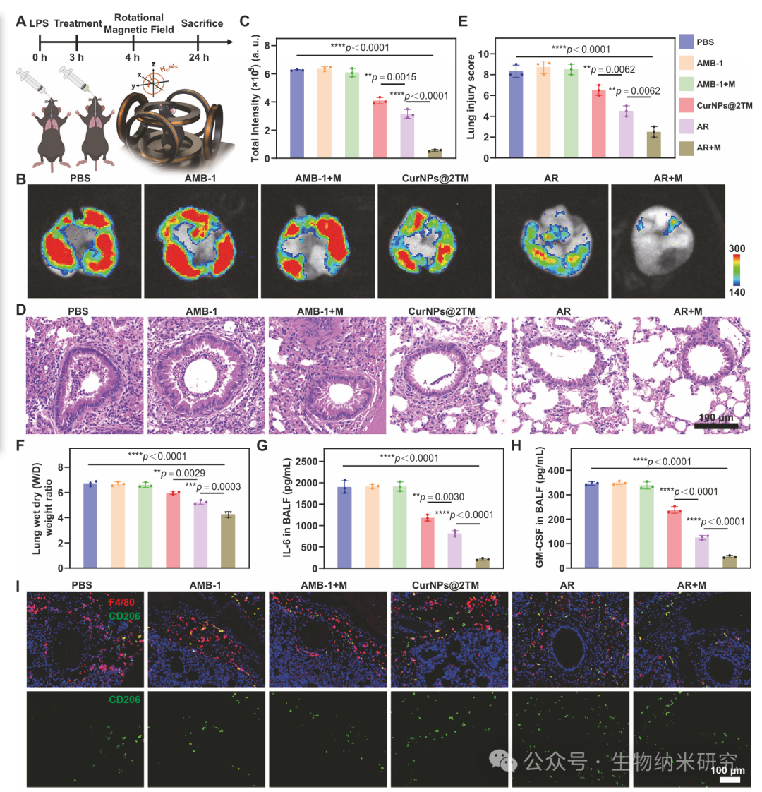
Figure 6 | AR Inhibits Acute Pneumonia
Click to read the original text, and you can jump to the original linkOthers are also reading Cooperation/Recommendation: TomHusky-