Competition Between Intrinsic Rhythm and Pacemaker Rhythm Leading to Misdiagnosis of Pacemaker Dysfunction

Competition Between Intrinsic Rhythm and Pacemaker Rhythm Leading to Misdiagnosis of Pacemaker Dysfunction

Competition Between Intrinsic Rhythm and Pacemaker Rhythm Leading to Misdiagnosis of Pacemaker Dysfunction

Guo Xiaoyu, Fan Xiaohan, Fan Shuxin

Wangjing Hospital, China Academy of Chinese Medical Sciences

Fuwai Hospital, Chinese Academy of Medical Sciences
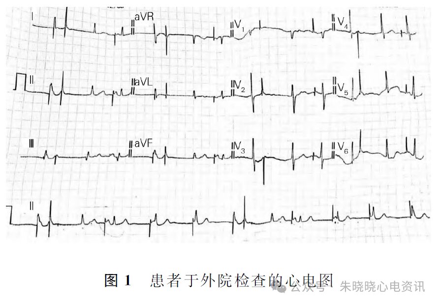
A 55-year-old female patient visited Fuwai Hospital on February 21, 2024, due to suspected pacemaker dysfunction after an ECG examination at another hospital.
The patient had a history of sick sinus syndrome, junctional escape rhythm, and frequent atrial premature beats, and had an Abbott dual-chamber pacemaker (model: Zephyr TM XL DR 5826) implanted for over 9 years.
Subsequently, a pacemaker programming check was performed at Fuwai Hospital, which showed that both pacing and sensing functions were normal.
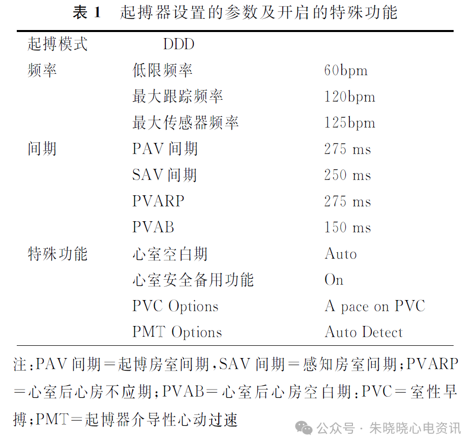
The ECG from the outside hospital is shown in Figure 1, with the pacemaker settings and functions listed in Table 1.

Competition Between Intrinsic Rhythm and Pacemaker Rhythm Leading to Misdiagnosis of Pacemaker Dysfunction

Competition Between Intrinsic Rhythm and Pacemaker Rhythm Leading to Misdiagnosis of Pacemaker Dysfunction

Analyzing the pacemaker function from the long II lead ECG (Figure 2). First, we analyze the atrial pacing function.
In the figure, both double pulses and single pulses can be seen, where ① and ⑤ are two sets of double pulses, and ②, ③, ④, ⑥, ⑦, ⑧, ⑨ are all single pulses.

Competition Between Intrinsic Rhythm and Pacemaker Rhythm Leading to Misdiagnosis of Pacemaker Dysfunction

R2 and R7 shapes are similar, without preceding P waves or pacing pulses, indicating junctional beats. Clear P waves can be seen after single pulses ② and ⑥, suggesting that these two single pulses are both A-P.
The intervals between pulses ②, ③, ④, ⑤, and between ⑥, ⑦, ⑧, ⑨ are all 1000 ms, consistent with the pacemaker’s lower limit frequency setting of 60 beats/min. Therefore, it is considered that single pulses ②, ③, ④, ⑥, ⑦, ⑧, ⑨ are all A-P, and the first pulse in double pulse ⑤ is also A-P. The interval of 275 ms between double pulses is consistent with the PA-V interval setting (see Table 1), suggesting AV sequential pacing (A-P – V-P), similarly for double pulse ①, which is also A-P – V-P.
After the A-P in the double pulse, pacing P waves can be seen merging with R1 and R6’s descending branches;
P waves can be seen after A-P ③ and ⑦, merging with R4 and R9 respectively;
A-P ④, ⑧, ⑨ are unrelated to the following R waves, with the subsequent R waves being slightly faster than the lower limit frequency of pacing, indicating accelerated junctional beats, consistent with the sources of R2 and R7 beats, but the following R waves are slightly different from the waveforms of R2 and R7, possibly due to the merging of A-P ④, ⑧, ⑨’s pacing P waves with the following junctional beats.
Thus, all A-Ps have pacing P waves, and no abnormalities were found in atrial pacing function.
Next, we analyze the atrial sensing function. The Abbott pacemaker timing method is an improved AA timing. During the intervals between A-P ②, ③, ④, ⑤, and ⑥, ⑦, ⑧, ⑨, R waves were not sensed during the VA intervals, and after the lower limit frequency interval of 1000 ms ended, A-Ps were sequentially discharged;
After double pulses ① and ⑤, R waves R2 and R7 were sensed during the VA intervals, restarting the pacing intervals from R2 and R7, but did not reach the lower limit frequency interval. Why were A-Ps ② and ⑥ discharged early?
Carefully examining the ascending branches of the T waves at R2 and R7, P waves (P1 and P2) can be seen, and the intervals between P1 and the subsequent A-P ②, and between P2 and the subsequent A-P ⑥ are equal, approximately 330 ms, suggesting that the A-Pace on PVC function is activated.
Checking the pacemaker settings, the A-Pace on PVC function was indeed activated (see Table 1).
Therefore, no abnormalities were found in the atrial sensing function either.
Now let’s analyze the ventricular pacing and sensing functions. In double pulses ① and ⑤, R1 and R6 are junctional beats, closely adjacent to the A-P in the double pulses, falling within the atrial post-ventricular blanking period (PAVB), and were not sensed by the ventricles, thus after the PAV interval ended, V-P was discharged, but V-P fell on the ascending branches of T waves R1 and R6, which are in the absolute refractory period of the ventricles, leading to functional loss of capture;
R3 and R8 are A-P paced junctional beats transmitted through the AV node, which were sensed by the ventricular channels, inhibiting the discharge of V-P;
R4, R5, R9, R10, and R11 are adjacent to A-P, unrelated to the preceding A-P, but without V-P afterwards, indicating that these QRS waves are detached from P-VAB, sensed by the ventricular channels, and did not cause cross-sensing. Such short A-P – VS intervals did not trigger the ventricular safety backup pulse, possibly related to the activation of the automatic ventricular blanking period.
Checking the pacemaker settings, the automatic ventricular blanking period function was indeed activated (see Table 1).
Thus, no abnormalities were found in the ventricular pacing and sensing functions.
Therefore, the diagnosis of this ECG is as follows:
1. Accelerated junctional escape rhythm + paced rhythm;
2. Dual-chamber pacemaker, DDD pacing mode;
3. No abnormalities in atrial pacing and sensing functions, and ventricular pacing and sensing functions;
4. The A-Pace on PVC function and automatic ventricular blanking period function are activated.
Discussion
The reason this ECG of the patient was misdiagnosed as pacemaker dysfunction is twofold:
First, the frequency of junctional escape beats is 60 bpm, and the pacemaker’s lower limit frequency is set at 60 bpm. Due to the close frequency, intrinsic beats and paced beats formed a competitive rhythm and fusion wave;
Second, the special functions of the pacemaker: A-Pace on PVC and automatic ventricular blanking period.
The A-Pace on PVC function is the Abbott pacemaker’s response to ventricular premature beats (VPBs). In this ECG, it is junctional escape beats, and the pacemaker senses that there are no preceding P waves and atrial pulses A-P, judging it as a VPB.
Upon determining that a VPB has occurred, the pacemaker actively extends the post-ventricular atrial refractory period (PVARP) to 480 ms, causing the subsequent P waves to fall within the refractory period and unable to continue triggering ventricular pacing, thus avoiding the occurrence of pacemaker-mediated tachycardia.
However, the position of the P waves after the premature beats varies, and the timing of subsequent atrial pacing discharges varies. In the extended 480 ms PVARP, the first 150 ms is the absolute refractory period (blanking period), and the following 330 ms is the relative refractory period. If there are no P waves after the VPB or the P waves fall within the blanking period, the pacemaker will operate normally with DDD timing.
If the P wave falls within the relative refractory period, it is defined as a retrograde P wave, and the pacemaker will discharge an atrial pacing pulse 330 ms after the retrograde P wave, followed by a ventricular pacing pulse after the programmed AV interval.
A-Pace on PVC can lead to significant advancement of atrial pacing, easily misinterpreted as impaired atrial sensing function.
The automatic ventricular blanking period (Auto Blanking) actually enhances the ventricular safety backup function, working by opening a 12 ms ventricular blanking period window after atrial pacing, followed by setting a refractory period window, not exceeding 52 ms.
If ventricular events are continuously sensed during the refractory window and reach the maximum limit of 52 ms, a 12 ms cross-sensing window (between 52 ms and 64 ms) is opened. If ventricular events are sensed during the cross-sensing window, a safety backup pulse will be discharged 120 ms after A-P.
If no ventricular events are sensed during the refractory period, or if events are sensed but terminate before the maximum limit of 52 ms, the cross-sensing window will not trigger, and it will directly enter an alert period.
The purpose of the automatic ventricular blanking period is to automatically extend the ventricular blanking period when cross-sensing exists, and maintain a shortened blanking period when cross-sensing does not exist, maximizing the reduction of inappropriate suppression caused by cross-sensing, thus maximizing the ventricular alert window.
The activation of the automatic ventricular blanking period in this case may lead to confusion when analyzing the ECG, as the QRS waves adjacent to the A-P did not trigger cross-sensing.
In summary, due to the competition between intrinsic rhythm and paced rhythm, and the operation of the pacemaker’s special functions, this ECG was misdiagnosed as pacemaker dysfunction. The lower limit frequency can be reduced through pacemaker programming to avoid the occurrence of competitive rhythms.
Moreover, when interpreting such paced ECGs, careful analysis of the relationship between paced beats and intrinsic beats should be conducted, first ruling out the possibility of activating special functions to avoid misdiagnosis and causing unnecessary psychological burdens to patients.

Competition Between Intrinsic Rhythm and Pacemaker Rhythm Leading to Misdiagnosis of Pacemaker Dysfunction

Competition Between Intrinsic Rhythm and Pacemaker Rhythm Leading to Misdiagnosis of Pacemaker Dysfunction

Leave a Comment