
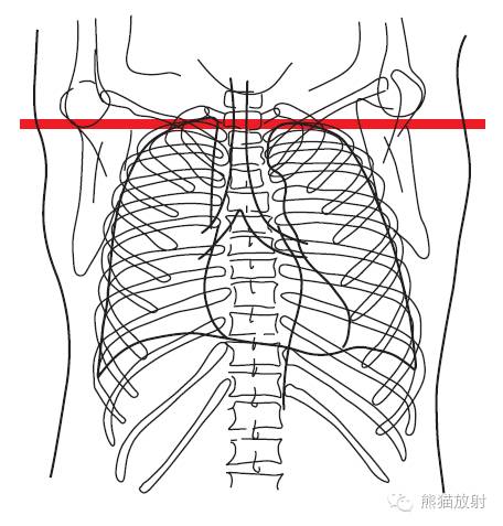
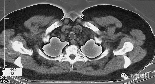
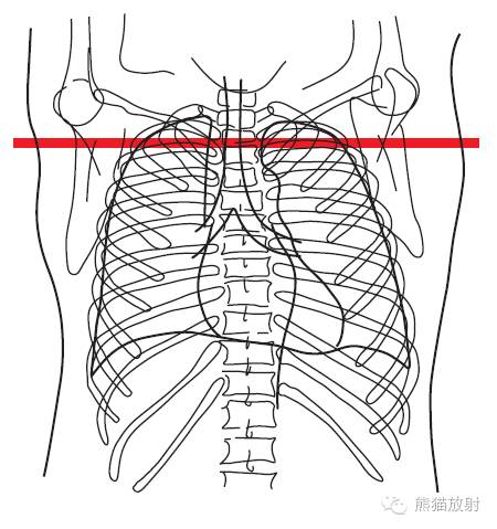
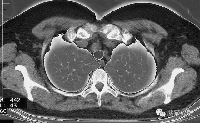
From the level of the thoracic inlet to the main pulmonary artery window, and from below the tracheal bifurcation to the level of the hepatic vein, each layer includes the positioning plane, axial CT images, corresponding schematic diagrams, anatomical structure names, simplified diagrams, lymph nodes, and pulmonary segment names. It’s incredibly comprehensive!
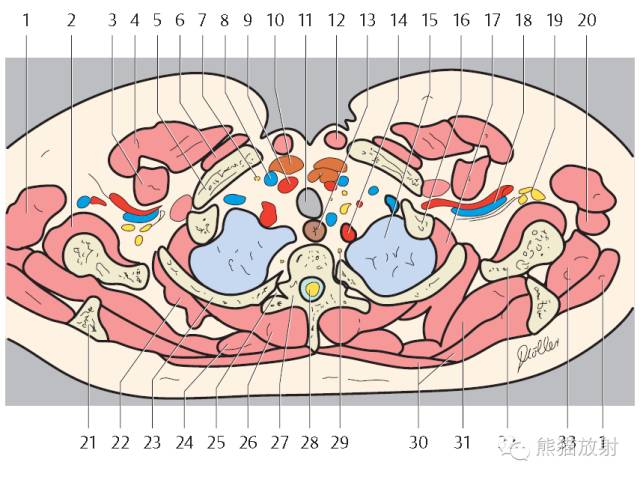
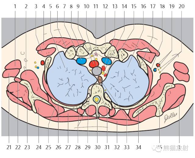
First Layer: Thoracic Inlet Level
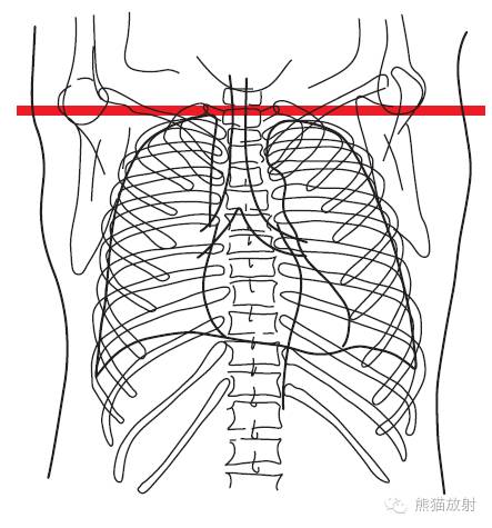
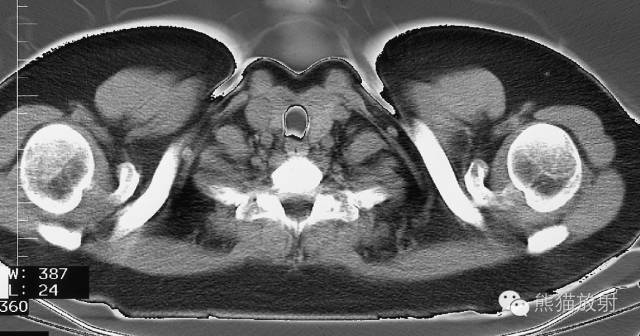
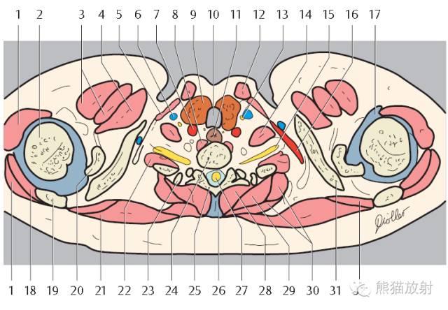
1. Deltoid muscle
2. Head of humerus
3. Pectoralis major muscle
4. Pectoralis minor muscle
5. Sternothyroid muscle
6. Internal jugular vein
7. Internal carotid artery
8. Longus colli muscle
9. Thyroid gland
10. Trachea
11. Esophagus
12. Sternocleidomastoid muscle
13. Vagus nerve (ramus)
14. Anterior scalene muscle
15. Brachial plexus
16. Subclavian artery
17. Joint capsule with capsular ligaments
18. Infraspinatus muscle
19. Spine of scapula
20. Coracoid process
21. Trapezius muscle
22. Medial and posterior scalenus muscles
23. Costovertebral joint (Rib 1)
24. Thoracic vertebra 1
25. Vertebral arch (lamina)
26. Supraspinous ligament
27. Spinal cord
28. Erector spinae muscle
29. Serratus anterior muscle
30. Rhomboid muscle
31. Clavicle
32. Supraspinatus muscle
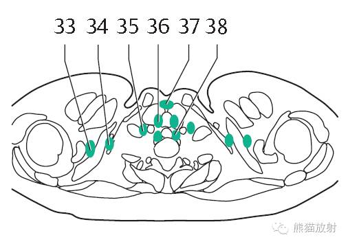
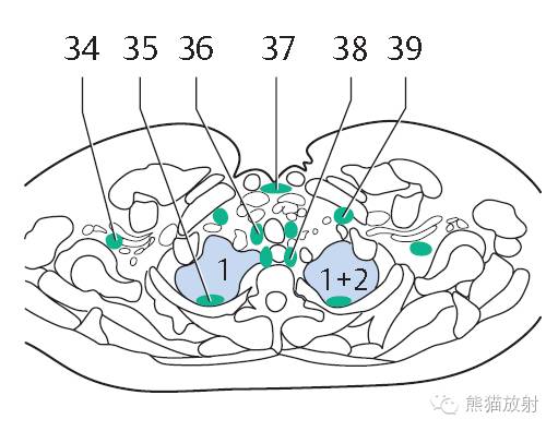
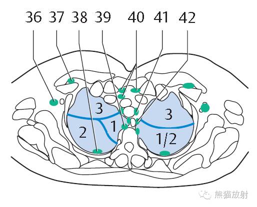
33. Supraclavicular lymph nodes
34. Subclavicular lymph nodes
35. Jugular lymph nodes
36. Paratracheal lymph nodes
37. Anterior cervical lymph nodes
38. Paraesophageal lymph nodes
Second Layer: Subclavian Artery and Vein Level
 1. Deltoid muscle
2. Subscapularis muscle
3. Pectoralis minor muscle
4. Pectoralis major muscle
5. Clavicle
6. Subclavius muscle
7. Vagus nerve (ramus)
8. Internal jugular vein
9. Internal carotid artery
10. Thyroid gland
11. Trachea
12. Sternocleidomastoid muscle
13. Esophagus
14. Subclavian artery
15. Left lung
16. Rib 1
17. Intercostal muscle
18. Subclavian artery and vein
19. Brachial plexus
20. Teres major muscle and latissimus dorsi muscle
21. Spine of scapula
22. Serratus anterior muscle
23. Rib 2
24. Rhomboid muscle
25. Costovertebral joint
26. Erector spinae muscle
27. Thoracic vertebra 2
28. Spinal cord
29. Sympathetic trunk
30. Trapezius muscle
31. Scapula
32. Infraspinatus muscle
34. Deep axillary lymph nodes
35. Intercostal lymph nodes
36. Paratracheal lymph nodes
37. Anterior cervical lymph nodes
38. Paraesophageal lymph nodes
Right Lung
1. Apical segment of upper lobe
Left Lung
1+2 Apicoposterior segment of upper lobe
Third Layer: Aortic Arch Three Major Branches Level
1. Deltoid muscle
2. Subscapularis muscle
3. Pectoralis minor muscle
4. Pectoralis major muscle
5. Right lung
6. Rib 1
7. Brachiocephalic vein
8. Clavicle
9. Brachiocephalic artery
10. Trachea
11. Thymus
12. Sternum (manubrium)
13. Left common carotid artery
14. Recurrent laryngeal nerve
15. Left subclavian artery
16. Left lung
17. Rib 2
18. Intercostal muscle
19. Axillary artery and vein
20. Brachial plexus
21. Teres major muscle and latissimus dorsi muscle
22. Spine of scapula
23. Serratus anterior muscle
24. Rib 3
25. Rhomboid muscle
26. Erector spinae muscle
27. Thoracic vertebra 2
28. Spinal cord
29. Sympathetic trunk
30. Trapezius muscle
31. Scapula
32. Infraspinatus muscle
33. Deep axillary lymph nodes
34. Interpectoral lymph nodes
35. Intercostal lymph nodes
36. Paratracheal lymph nodes
37. Anterior cervical lymph nodes
38. Paraesophageal lymph nodes
39. Jugular lymph nodes
Right Lung
1. Apical segment of upper lobe
右肺上叶尖段
Left Lung
1+2 Apicoposterior segment of upper lobe
左肺上叶尖后段
Fourth Layer: Aortic Arch Upper Edge Level
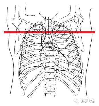
1. Deltoid muscle
2. Subscapularis muscle
3. Pectoralis minor muscle
4. Pectoralis major muscle
5. Right lung
6. Rib
7. Right brachiocephalic vein
8. Right brachiocephalic artery
9. Vagus nerve
10. Trachea
11. Thymus
12. Sternum (manubrium)
13. Left common carotid artery
14. Recurrent laryngeal nerve
15. Left subclavian artery
16. Left lung
17. Rib 2
18. Intercostal muscle
19. Axillary artery and vein
20. Brachial plexus
21. Teres major muscle and latissimus dorsi muscle
22. Spine of scapula
23. Serratus anterior muscle
24. Rib 3
25. Rhomboid muscle
26. Erector spinae muscle
27. Thoracic vertebra
28. Spinal cord
29. Sympathetic trunk
30. Trapezius muscle
31. Scapula
32. Infraspinatus muscle
33. Deep axillary lymph nodes
34. Interpectoral lymph nodes
35. Intercostal lymph nodes
36. Paratracheal lymph nodes
37. Anterior mediastinal lymph nodes
38. Paraesophageal lymph nodes
39. Jugular lymph nodes
Right Lung
4. Lateral segment of middle lobe
中叶外侧段
5. Medial segment of middle lobe
中叶内侧段
6. Superior segment of lower lobe
下叶上段
Left Lung
4. Superior lingular segment
上舌段
5. Inferior lingular segment
下舌段
6. Superior segment of lower lobe
下叶上段
10. Posterior basal segment of lower lobe
下叶后基底段
Fifth Layer: Pulmonary Artery Trunk Level
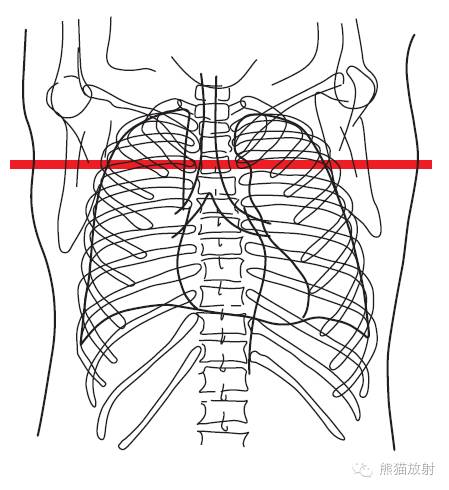
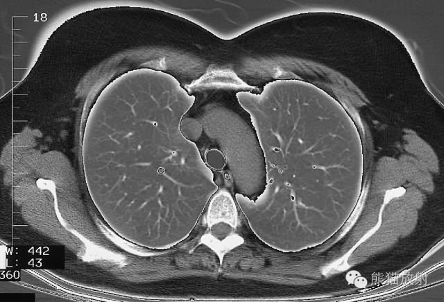
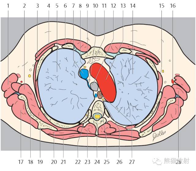 1. Latissimus dorsi muscle
2. Subscapularis muscle
3. Pectoralis minor muscle
4. Pectoralis major muscle
5. Right lung
6. Rib
7. Right pulmonary artery
8. Phrenic nerve
9. Sternum
10. Right atrium
11. Left lung
12. Intercostal muscle
13. Thoracic vertebra
14. Spinal cord
15. Descending aorta
16. Esophagus
17. Trachea
18. Right main stem bronchus
19. Left main stem bronchus
20. Right inferior lobar bronchus
21. Left inferior lobar bronchus
22. Right lung
23. Left lung
24. Azygos vein
25. Thoracic duct
26. Vagus nerve
27. Descending aorta
28. Right pulmonary vein
29. Left pulmonary vein
30. Azygos vein
31. Right main stem bronchus
32. Left main stem bronchus
33. Bronchopulmonary lymph nodes
34. Intercostal lymph nodes
35. Paravertebral lymph nodes
36. Parasternal lymph nodes
37. Juxtaesophageal lymph nodes
38. Para-aortal lymph nodes
39. Intercostal lymph nodes
40. Right lung
41. Left lung
42. Azygos vein
43. Thoracic duct
44. Vagus nerve
45. Descending aorta
46. Right main stem bronchus
47. Left main stem bronchus
48. Right inferior lobar bronchus
49. Left inferior lobar bronchus
50. Right lung
51. Left lung
来源:熊猫放射整理