Recently, the expert team at our hospital’s Gynecological Oncology Center successfully utilized a 3D printed individualized navigation template to guide the Large Segment Implant Radiation Therapy (HIBT) technique, precisely treating a patient with a large and complex metastatic lesion in the sacral region due to uterine sarcoma. This innovative application, which integrates cutting-edge guidance technology with an efficient radiation therapy model, opens new pathways for managing such challenging metastatic lesions.
3D Printing Precision Navigation
The Guiding Light of Large Segment Radiation Therapy
Two years ago, Ms. Li (pseudonym) was diagnosed with highly malignant uterine leiomyosarcoma (ULMS). Despite multiple treatments, her condition continued to progress. Two months ago, she developed metastases in the sacral region, with a large and irregularly shaped lesion closely adjacent to the spinal cord and blood vessels, causing severe pain and presenting significant challenges for traditional treatment.
In response to the complex anatomical characteristics of the lesion, the team led by Dr. Chen Wenjuan at the Gynecological Oncology Center collaborated with Dr. Chen Juhui’s team from the Department of Abdominal Tumor Radiation Therapy and the physics team to innovatively integrate 3D printing technology into the HIBT process:
1
Precise Modeling
Integrating multimodal imaging such as CT and MRI to accurately reconstruct the metastatic lesion and adjacent critical structures (spinal cord, blood vessels).
2
Individualized Template
Using 3D printing technology, a personalized navigation template that perfectly fits the patient’s anatomical structure was custom-designed, carefully planning and pre-setting the optimal spatial layout and puncture path for the implantation needle.
3
Precise Guidance for Implantation
Under the real-time, visual guidance of the 3D printed template, the implantation needle was accurately and safely placed into the target position.
4
Large Segment Enhancement
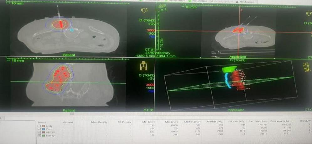
Combining an optimized large segment radiation therapy plan (single dose > 10 Gy), achieving high-dose, high-precision irradiation of the lesion while maximizing protection of surrounding normal tissues.
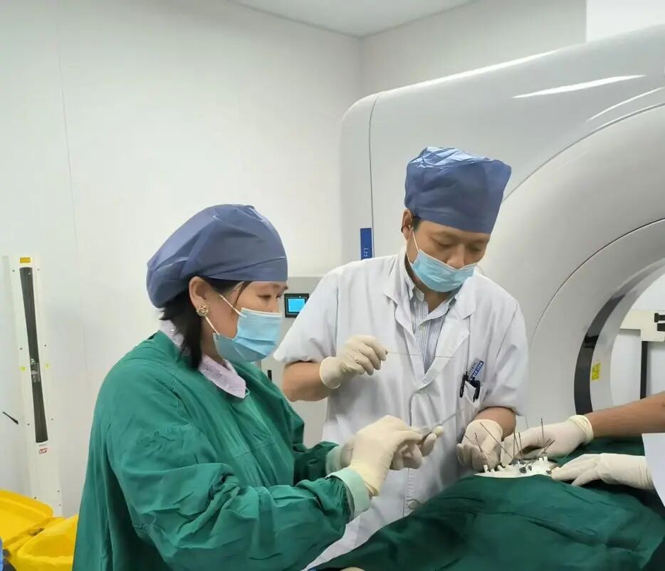
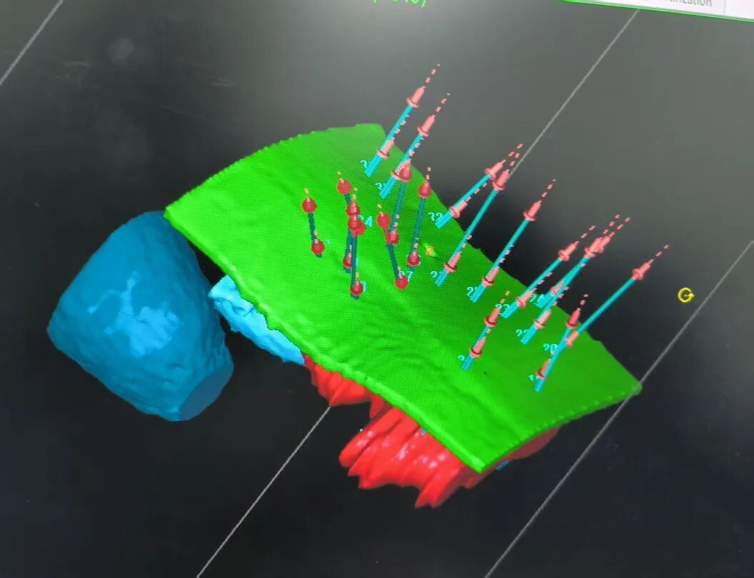
Thanks to the precise positioning guided by 3D printing and the efficient treatment of the large segment mode, Ms. Li’s pain symptoms were significantly alleviated after treatment.
Advantages of Technology Integration
Breakthroughs in Precision and Efficiency
This successful case highlights the core advantages of 3D printing technology guiding HIBT:
1
Revolution in Spatial Precision
The individualized 3D printed template addresses the challenges of manual implantation in complex anatomical areas (such as the sacral region), ensuring the precise placement of the radiation source, which is a prerequisite for safely implementing high-dose radiation therapy.
2
Guarantee of Efficient Treatment
The precise implantation combined with the large segment mode (high single dose, short treatment course of 1-2 weeks) can more effectively overcome the radiation resistance of uterine leiomyosarcoma, significantly enhancing local control potential and improving patient tolerance.
3
New Hope for Complex Cases
Provides a powerful precision treatment option for isolated or few metastatic lesions that are large, irregular, and adjacent to critical organs, which are difficult to manage with traditional surgery or external radiation.
Broad Clinical Application Prospects
Dr. Chen Wenjuan emphasized that the 3D printing-guided large segment implantation radiation therapy technology not only successfully achieved precise targeting of this complex metastatic lesion but also represents our center’s cutting-edge exploration in the field of precise treatment for gynecological tumors. This technology integrates the dual advantages of spatial precision guidance (3D printing) and efficient biological effects (large segment), with broad clinical application prospects:
1
Promotion to More Complex Sites
Such as pelvic recurrence lesions, head and neck tumors, and other areas that are difficult to cover with surgery or external radiation.
2
Enhancing Efficacy for Refractory Tumors
Providing more effective local treatment options for tumors with strong radiation resistance and difficult locations (such as ULMS).
3
Optimizing Diagnosis and Treatment Processes
The repeatability and precision of the 3D printed template help standardize operations, shorten the learning curve, and are expected to benefit more patients in the future.
In the future, the Gynecological Oncology Center will continue to deepen the exploration and promotion of this technology, committed to providing more precise and efficient treatment options for cancer patients.
Gynecological Oncology Center
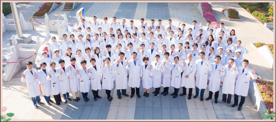
The Gynecological Oncology Center of Fujian Cancer Hospital is a key clinical specialty in Fujian Province, a clinical medical research center for precise treatment of gynecological malignant tumors, and a training point for master’s and doctoral students from Fujian Medical University and Fuzhou University. It is one of the first national pilot centers for quality control of standardized diagnosis and treatment of ovarian and cervical cancer, and serves as the chair unit of the Gynecological Oncology Branch of the Fujian Medical Association and the Gynecological Oncology Committee of the Fujian Anti-Cancer Association. It is also currently the largest comprehensive treatment center for gynecological tumors in the province, with 4 wards, 220 beds, and the highest number of cases treated annually for cervical cancer, ovarian cancer, endometrial cancer, and vulvar cancer among major hospitals in the province. In recent years, while maintaining high-quality clinical work, the center has improved the quality and efficiency of scientific research, continuously obtaining funding for multiple important projects, especially achieving new results in clinical translational research. Currently, the center undertakes numerous international and national multi-center clinical research projects, striving to benefit a wide range of cancer patients with the latest scientific achievements. In the future, the department will focus more on the collaborative development of clinical and scientific research, building a research-oriented and innovative gynecological oncology treatment center to contribute to the construction of national regional cancer medical centers.
Gynecological Oncology Center / Contribution
Feng Mei / Review
 Editing | Fu Mengying, Lin Guihua