
In July 2025, Professor Ann M Parr’s team at the University of Minnesota published a high-level research paper titled: 3D-Printed Scaffolds Promote Enhanced Spinal Organoid Formation for Use in Spinal Cord Injury in the journal Advanced Healthcare Materials (IF: 9.6).
Abstract
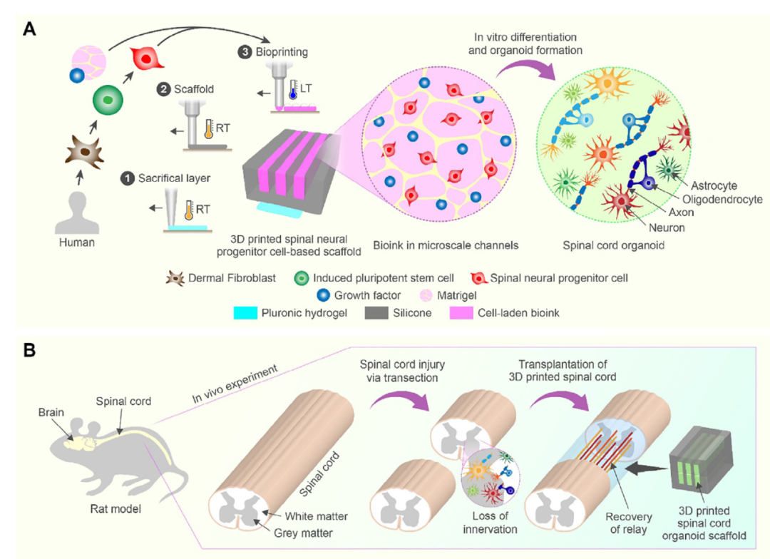
Transplantation of region-specific spinal neural progenitor cells (sNPCs) has shown promise in restoring function after spinal cord injury (SCI) by forming connections with host neural circuits. Here, a 3D-printed organoid scaffold was developed for the transplantation of clinically relevant human induced pluripotent stem cell-derived region-specific sNPCs.
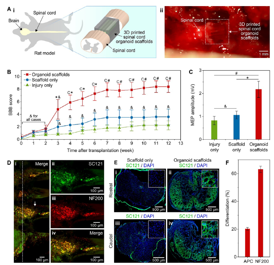
The scaffold was printed with micro-scale channels, followed by the printing of sNPCs within these channels. The scaffold guides axonal projections along the channels and simulates in vivo-like conditions for more effective cell maturation and neuronal network development, which is crucial for functional recovery after SCI. The scaffold and organoids assembled along its length were transplanted into the transected spinal cord of rats, significantly promoting functional recovery. After 12 weeks post-transplantation, most cells within the scaffold differentiated into neurons and integrated into the host spinal cord tissue.
These results demonstrate their potential to create relay systems along the spinal cord and form synapses in the rostral and caudal directions relative to the scaffold. The combination of sNPCs, organoid assembly, and 3D printing strategies is expected to lead to transformative therapeutic approaches for SCI.
Innovations
1. A novel method for treating spinal cord injury (SCI) was developed by combining multi-material 3D printing technology with human induced pluripotent stem cell (iPSC)-derived region-specific spinal neural progenitor cells (sNPCs) to create 3D-printed spinal organoid scaffolds with microchannels.
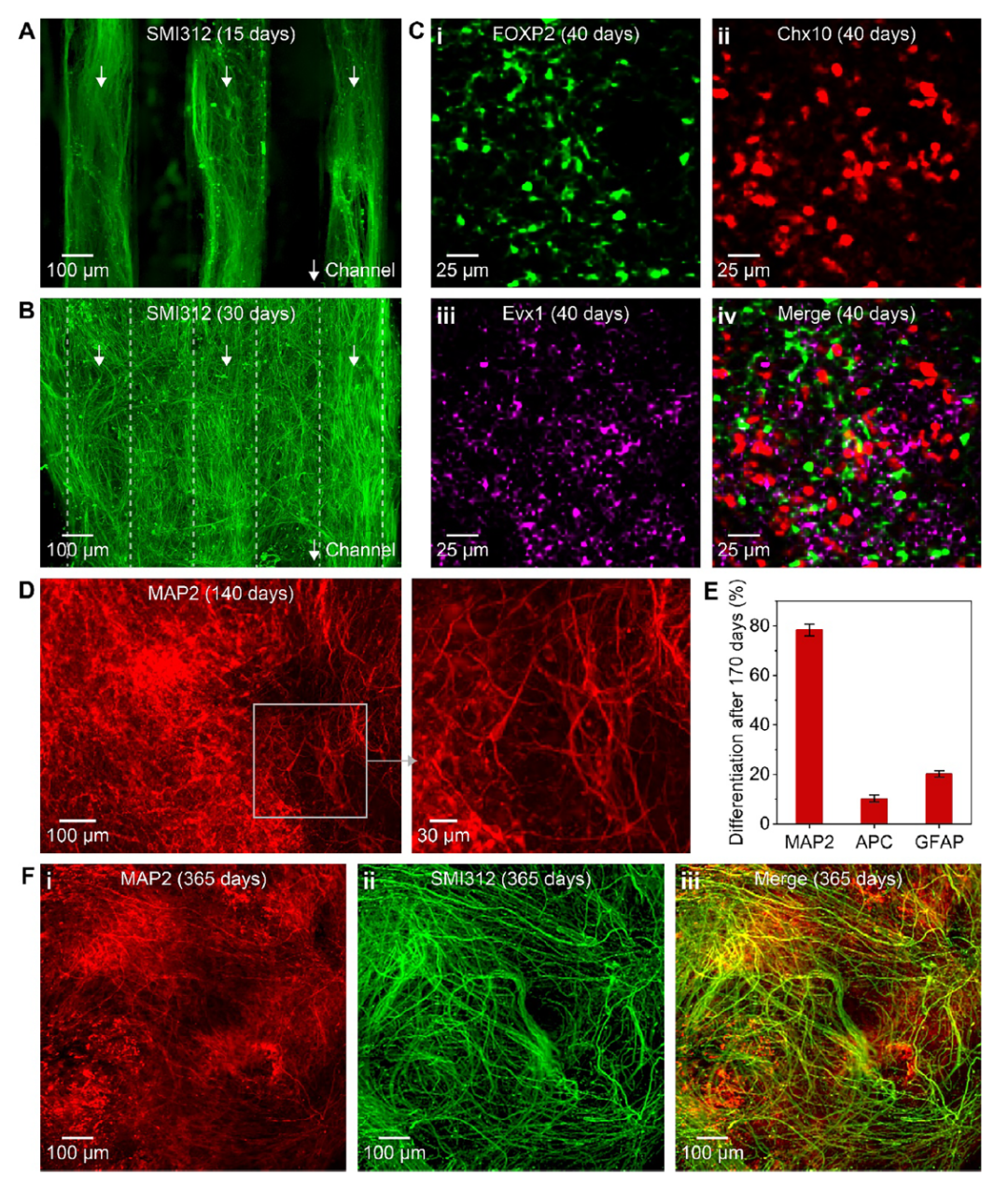
2. The 3D-printed sNPCs differentiated into various spinal-specific cell types within the scaffold, forming a layered structure resembling spinal tissue and maintaining neuronal characteristics in vitro for at least one year, exhibiting highly organized axon bundles.
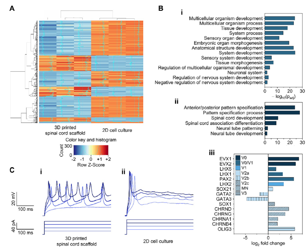
3. RNA sequencing and electrophysiological experiments confirmed that the gene expression of 3D-printed sNPCs is closer to spinal tissue, showing higher expression levels of genes related to spinal regionalization and neuronal maturation. Functionally, they exhibited more mature neuronal characteristics compared to 2D cultured cells, with higher maturity and functionality.
4. Transplantation of the 3D-printed spinal organoid scaffold into a complete transection rat model significantly promoted functional recovery, including improvements in motor function and enhanced nerve conduction.
5. The transplanted scaffold induced axons and dendrites to extend at the injury site, forming functional synaptic connections with the host spinal tissue, demonstrating the scaffold’s potential in promoting nerve regeneration and integration.
Q&A
Q1: What are the main advantages of the 3D-printed spinal organoid scaffold?
A: The main advantage of the 3D-printed spinal organoid scaffold is its ability to provide precise mechanical guidance and structural support for spinal neural progenitor cells (sNPCs). This scaffold guides axonal growth through microchannels, promoting cell maturation and neural network formation, thereby better simulating the physiological environment of the spinal cord. Additionally, 3D printing technology allows for customization of the scaffold according to the shape of the injury site, improving integration post-transplantation and providing a more effective strategy for spinal injury repair.
Q2: What are the significant differences between 3D-printed sNPCs and 2D cultured sNPCs in vitro?
A: The 3D-printed sNPCs exhibit gene expression that is closer to spinal tissue, particularly showing higher expression levels of genes related to spinal regionalization and neuronal maturation. Electrophysiological experiments indicate that neurons differentiated from 3D-printed sNPCs possess more mature electrophysiological characteristics, capable of generating continuous repetitive action potentials, which 2D cultured cells cannot achieve. Furthermore, 3D-printed sNPCs can maintain neuronal characteristics in vitro for at least one year, displaying highly organized axon bundles, while 2D cultured cells fail to achieve such long-term stability and organization.
Q3: How does the 3D-printed spinal organoid scaffold promote functional recovery in rats in vivo?
A: The scaffold provides structural support at the injury site, preventing cells from drifting or aggregating post-transplantation, thus enhancing cell survival rates. Additionally, the sNPCs within the scaffold differentiate into various spinal-specific cell types, including neurons and oligodendrocytes, which can extend axons and dendrites, forming functional connections with the host spinal tissue. Moreover, measurements using the Basso, Beattie, and Bresnahan (BBB) motor function scoring and motor-evoked potentials (MEPs) show that rats receiving the 3D-printed spinal organoid scaffold transplantation exhibit significant recovery in motor function and enhanced nerve conduction, indicating that the scaffold can promote nerve regeneration and functional recovery.
Results
Original Article Link
Original link:
https://pubmed.ncbi.nlm.nih.gov/40702833/

Copyright Statement
The label ‘original’ only represents original translation. This platform does not claim copyright over the original text. The platform’s reprint is solely for academic exchange and information dissemination, and does not represent the platform’s views or verify the authenticity of its content. The copyright of the original text belongs to the original author. If the author does not wish to be reprinted or has any infringement, please contact this platform for deletion.