Robert G. Briggs and colleagues from the Department of Neurosurgery at the University of Southern California in Los Angeles studied the Superior Frontal Gyrus (SFG) and its white matter connections through MRI imaging of brain fiber bundles in healthy adults and anatomical studies of cadaver brains. Their results were published online in the journal Clin Anat in November 2019.
——Excerpt from the article chapter

Long press the QR code or click “Read the original text” to view the original text

[Ref: Briggs RG, et al. Clin Anat. 2019 Nov 21. doi: 10.1002/ca.23523. [Epub ahead of print]]
Research Background
The Superior Frontal Gyrus (SFG) is an important area that controls multiple functions such as movement, working memory, resting state, and cognition. Anatomical studies of different parts of the SFG can help better understand the significance of the SFG in human cognitive functions and the consequences of tumor surgery. Robert G. Briggs and colleagues from the Department of Neurosurgery at the University of Southern California in Los Angeles studied the SFG and its white matter connections through MRI imaging of brain fiber bundles in healthy adults and anatomical studies of cadaver brains. Their results were published online in the journal Clin Anat in November 2019.
Research Methods
This study used diffusion spectrum imaging (DSI) data from 10 healthy adults’ brains from the Human Connectome Project to perform fiber bundle imaging, assessing the connections of the SFG with other brain regions as a whole. Based on the relationship between the white matter fiber bundles shown by DSI and surrounding important neuroanatomical structures, the study characterized the features of the SFG fiber bundles. All fiber bundles were located in the bilateral cerebral hemispheres, and their lateralization indices were calculated based on fiber bundle volumes. A modified Klingler technique was used for microscopic anatomical studies of 10 cadaver brains to determine the locations of the main connecting fiber bundles of the SFG. The researchers divided the cortical surface of the SFG into an upper lateral side and a medial side. The frontal pole is the anterior edge of the SFG, while the central sulcus and the paracentral lobule are the posterior edges of the SFG (Figure 1).
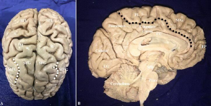
Figure 1. Anterior superior view (A) and mid-sagittal view (B) of the brain, showing parts of the frontal, parietal, and occipital lobes. In Figure A, the superior frontal sulcus is delineated by white dots; in Figure B, the cingulate sulcus is delineated by black dots; the lateral and medial sides of the SFG are clearly visible. CC: Corpus callosum; FP: Frontal pole; LG: Lingual gyrus; M: Motor center; MB: Midbrain; MFG: Middle frontal gyrus; PL: Paracentral lobule; S: Sensory center; SFG: Superior frontal gyrus; SPL: Superior parietal lobule.
The researchers ultimately identified four fiber bundles connected to the SFG: the Frontal Aslant Tract (FAT) connecting the inferior frontal gyrus; the Inferior Fronto-Occipital Fasciculus (IFOF) connecting the cuneus, lingual gyrus, and superior parietal lobule; the Cingulum connecting the precuneus, parahippocampal gyrus/hook; and the callosal fibers connecting the bilateral SFG.
The FAT connects the SFG and the inferior frontal gyrus in the same hemisphere, originating from the upper lateral surface of the SFG just anterior to the precentral gyrus, descending into the deep white matter of the frontal lobe, and then gradually bending approximately 90 degrees posterior-inferiorly, terminating in the opercular and triangular parts of the inferior frontal gyrus (Figure 2). The extent of the FAT extending along the SFG varies among subjects, but no origin of the FAT was found in the frontal pole, nor was it found to originate from the medial cortex of the SFG. It is important to note that the FAT runs deep within the superior longitudinal fasciculus, and carefully removing the superior longitudinal fasciculus is crucial for exposing the FAT in the deep white matter.
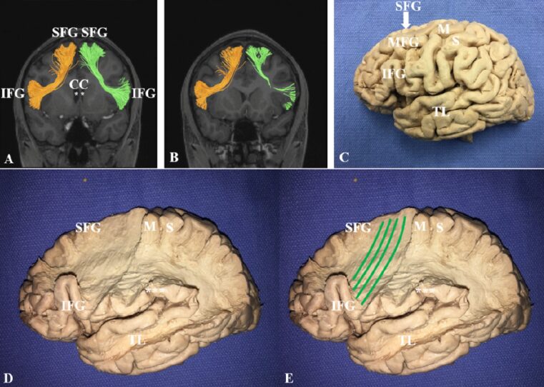
Figure 2. Frontal Aslant Tract. A, B: MRI coronal view showing FAT fiber bundle images. C: Lateral structure of the frontal lobe. D, E: FAT from the posterior lateral part of the SFG to the opercular and triangular parts of the inferior frontal gyrus (IFG); the green streamlines in Figure E show the trajectory of the FAT fibers. *: Lateral ventricle; CC: Corpus callosum; IFG: Inferior frontal gyrus; M: Motor center; MFG: Middle frontal gyrus; S: Sensory center; SFG: Superior frontal gyrus; TL: Temporal lobe.
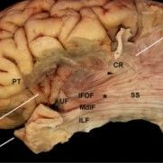
The Inferior Fronto-Occipital Fasciculus (IFOF) originates from the upper lateral SFG, converging in the white matter beneath the insula anterior to the external capsule and posterior to the outer capsule. Before dispersing in the deep white matter of the temporal lobe, the fiber bundle travels down laterally to the thalamus. The dispersed IFOF fibers continue projecting posteriorly and superiorly, approaching the lateral ventricle before terminating in the parietal and occipital lobes (Figure 3).
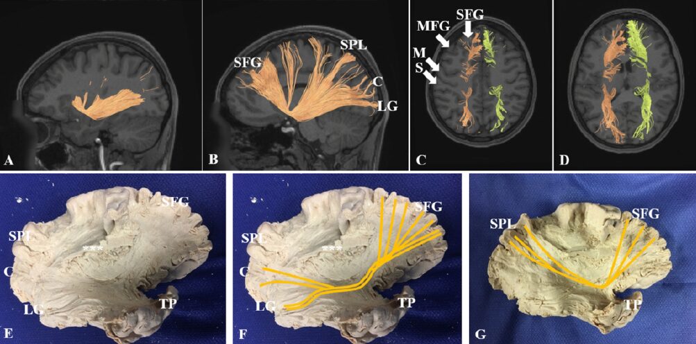
Figure 3. MRI-T1 weighted images showing the fiber bundle imaging of the Inferior Fronto-Occipital Fasciculus in sagittal (A, B) and axial (C, D) views. The IFOF travels longitudinally along the SFG before dispersing in the deep white matter of the posterior temporal lobe, terminating in parts of the parietal and occipital lobes. E, F: IFOF extending from the SFG to the cuneus and lingual gyrus; the orange streamlines in Figure F show the trajectory of the IFOF fibers. G: After removing the remaining cortical tissue in Figure F (***), the IFOF can be seen terminating in the superior parietal lobule; the orange streamlines in Figure G show the trajectory of the IFOF fibers. C: Cuneus; LG: Lingual gyrus; M: Motor center; MFG: Middle frontal gyrus; S: Sensory center; SFG: Superior frontal gyrus; SPL: Superior parietal lobule; TP: Temporal pole.
The Cingulum is located on the medial side of the SFG and is divided into anterior, middle, and posterior parts. The fibers of the cingulate gyrus curve 90 degrees posteriorly deep within the cingulate gyrus, with most fibers projecting upward and inward, terminating in the precuneus. However, part of the cingulum that curves 180 degrees continues posteriorly after the posterior part of the corpus callosum, projecting into the medial deep temporal lobe. The remaining fibers terminate in the parahippocampal gyrus and the hook (Figure 4).
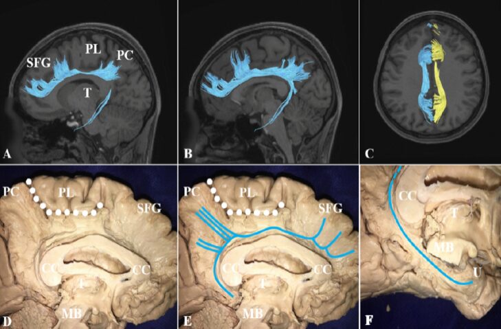
Figure 4. MRI-T1 weighted images showing the fiber bundle imaging of the Cingulum in sagittal (A, B) and axial (C) views. D, E: Cingulum extending from the medial side of the SFG to the precuneus; some cingulum fibers can be seen projecting posteriorly around the posterior part of the corpus callosum, terminating in the parahippocampal gyrus and the hook. In Figure F, after removing the midbrain, the cingulum can be seen entering the medial temporal lobe. The white dots in Figures D and E delineate the boundaries of the cingulate sulcus, and the light blue streamlines in Figures E and F show the trajectory of the cingulum fibers. CC: Corpus callosum; MB: Midbrain; PC: Precuneus; PL: Paracentral lobule; SFG: Superior frontal gyrus; T: Thalamus; U: Hook.
The SFG on the left and right hemispheres is connected by dense fibers of the corpus callosum. The projecting fibers of the corpus callosum connect the bilateral SFG through the knee and body of the corpus callosum (Figure 5). Most of the fibers projecting from the corpus callosum to the frontal pole travel horizontally. Most of the posterior corpus callosum fibers originate from the supplementary motor area (SMA), projecting vertically through the anterior part of the corpus callosum, connecting homologous regions of the bilateral SFG.
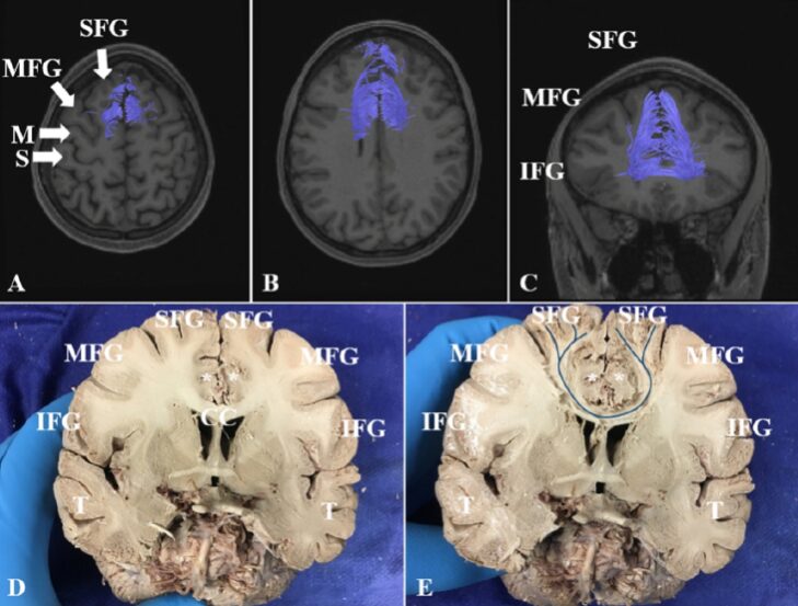
Figure 5. MRI-T1 weighted images showing the axial (A, B) and coronal (C) fiber bundle imaging of the corpus callosum. D, E: Cadaver coronal anatomical images showing the corpus callosum. * indicates bilateral cingulate gyri. The dark blue streamlines in Figure E show the trajectory of the corpus callosum fibers. CC: Corpus callosum; IFG: Inferior frontal gyrus; MFG: Middle frontal gyrus; SFG: Superior frontal gyrus; T: Temporal lobe.
In addition to the aforementioned four major white matter fiber bundles, the researchers discovered many short U-shaped association fibers during the fiber bundle anatomical study (Figure 6). The U-shaped bundles mainly connect the SFG or the middle frontal gyrus, with each U-shaped bundle having a similar morphology, originating from a certain part of the cortex and terminating in adjacent cortex.
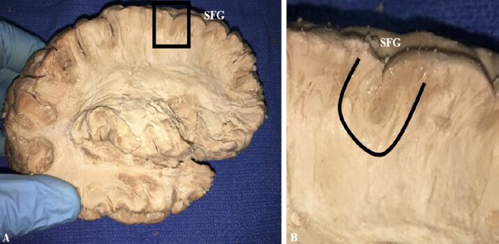
Figure 6. U-shaped bundles connecting adjacent cortex within the white matter of the SFG. The black box in Figure A shows the U-shaped bundles; Figure B is an enlarged view of the U-shaped bundles. SFG: Superior frontal gyrus.
Conclusion
Finally, the authors pointed out that the SFG is mainly involved in various important functions such as movement, working memory, and higher cognitive activities; the brain network systems including FAT, IFOF, cingulum, and corpus callosum connect various parts of the SFG with other brain regions to execute and coordinate these functions. Research on the anatomy of the SFG and its fiber bundle connections helps explain the reasons for neurological dysfunctions occurring after tumor surgeries in the SFG and surrounding areas.
Compiled by

Li Xinxiao, MD
Ningxia Medical University General Hospital
Translated by

Hu Kejia, MD
Shanghai Jiao Tong University School of Medicine
Ruijin Hospital Affiliated
Reviewed by

Li Xinxiao, MD
Ningxia Medical University General Hospital
Final Review

Chen Xiancheng, Professor
Editor-in-Chief of Neurosurgery Information
Huashan Hospital Affiliated to Fudan University