Introduction: Continuing with instrument-assisted diagnosis, today we encounter a transparent cast. In clinical practice, the most important type to recognize is the transparent cast. Other casts appear as contents within the “transparent cast”; for example, if particles are present, it is termed a granular cast, and if cells are present, it is termed a cellular cast. If the “transparent cast” appears waxy, it is referred to as a waxy cast, and so on. Today, we will examine the instrument-assisted diagnosis of transparent casts.
  PART01: Patient: Female, 62 years old, undergoing routine urinalysis for glomerulonephritis follow-up.★ The results of the urinalysis are as follows. There are two issues to note in the regular records: one is that the cellular deviation exceeds one gradient, and the other is the presence of visible transparent casts and pathological casts. In the result area in the middle, a large number of transparent casts can be seen.
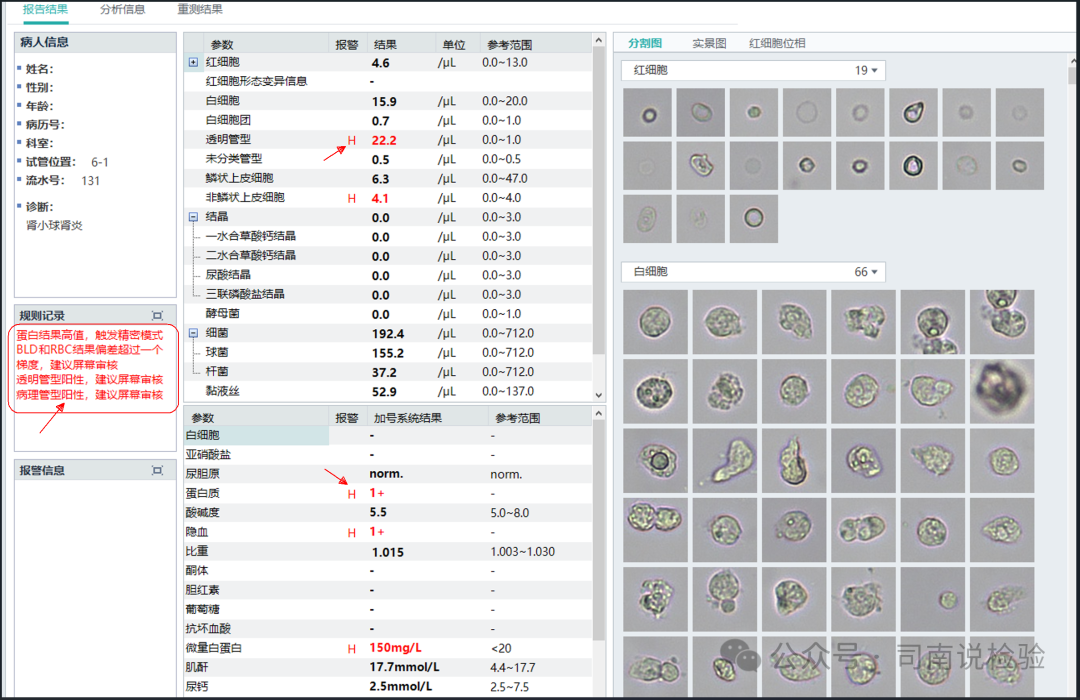 ★ Today, we will focus on the transparent cast section, enlarging this area:
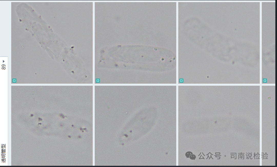 ★ Some colorless, transparent, thin-textured, poorly refractive, cylindrical transparent casts can be seen, with several enlarged.
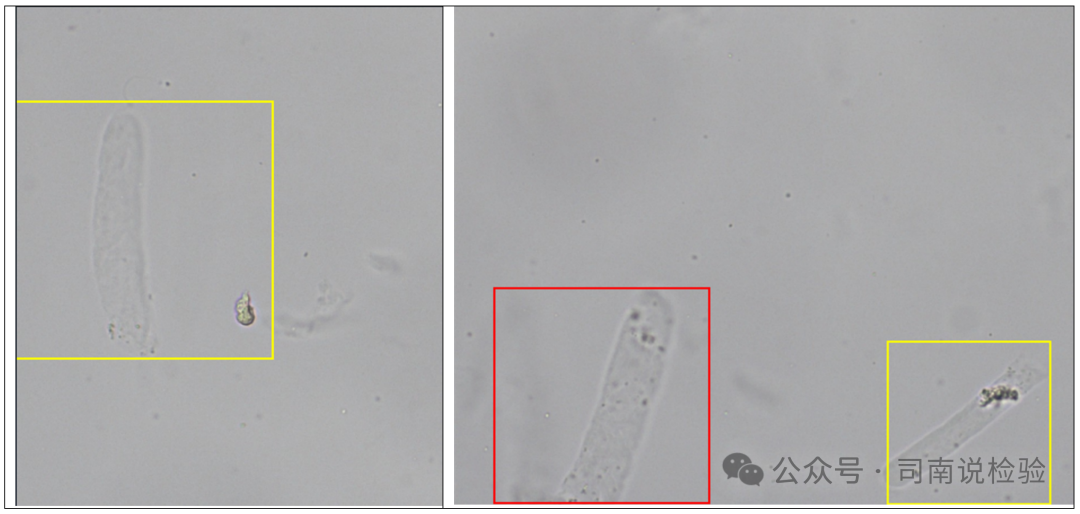 ★ The red box indicates the currently selected structure, while the yellow indicates other similar structures. Centrifugal microscopy:
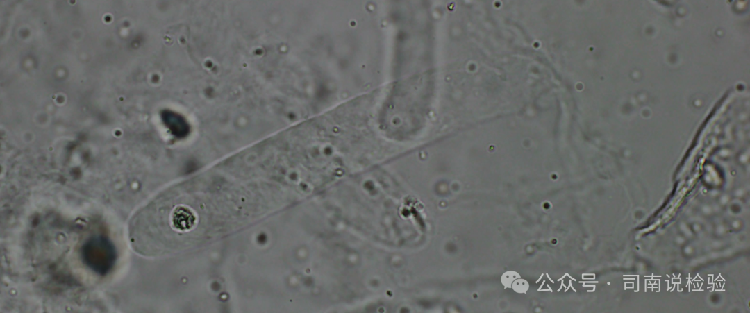 ★ Transparent casts, due to their minimal contents, need to be observed under dark field microscopy. It is important to note that under conditions where crystals (especially amorphous crystals), bacteria, or mucus threads are present, these substances can adhere to the transparent casts. At this time, it is necessary to adjust the fine focus screw to clarify the specific structure.
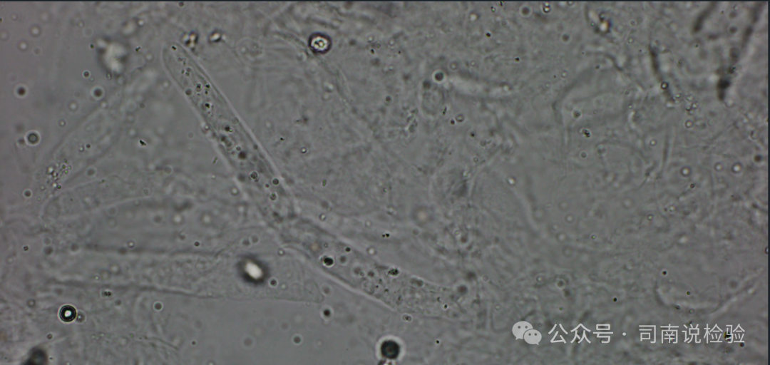 ★ It is important to note that transparent casts are not absolutely devoid of contents. According to expert consensus, a cast is only termed a “cast” when it contains more than 1/3 of its volume. In clinical practice, this is not a rigid rule; the occurrence and development of diseases do not have absolute stages. Early, middle, and late stages are artificially divided. Therefore, in clinical practice, one should base their understanding on theory and then dialectically view the disease. For instance, the term “normal result” does not imply that the absence of a single arrow indicates normality.
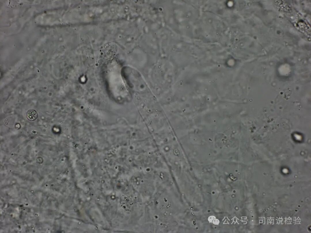 ★ It is crucial to differentiate transparent casts from mucus threads. Transparent casts are generally parallel on both sides, with clear edges and blunt round ends, while mucus threads are not. Their morphology varies widely, with different lengths, folds, twists, and irregular edges. However, transparent casts are not always perfectly straight at both ends; they can also exhibit folds, twists, and breaks. One should not interpret this based solely on textbook knowledge, as real situations are more complex than what is presented in textbooks. Another point to note is the correlation between casts and proteins; there are many cases where transparent casts appear with negative dry chemical protein results, so one should be cautious not to be misled by preconceived notions.
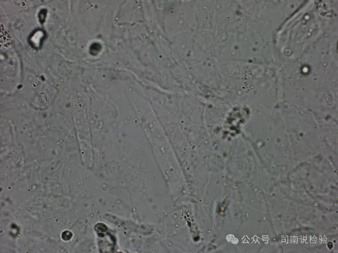 ★ Extended Knowledge Point: Urine – Casts – Transparent Casts
 Final Thoughts: Transparent casts are the most common type of casts. It is generally believed that transparent casts contain no substances; however, according to the latest interpretations, a cast can still be termed a transparent cast if its contents are less than 1/3. Transparent casts are typically parallel on both sides, with clear edges and blunt round ends, while mucus threads are not. Their morphology varies widely, with different lengths, folds, twists, and irregular edges. However, transparent casts are not always perfectly straight at both ends; they can also exhibit folds, twists, and breaks. One should not interpret this based solely on textbook knowledge, as real situations are more complex than what is presented in textbooks. Another point to note is the correlation between casts and proteins; there are many cases where transparent casts appear with negative dry chemical protein results, so one should be cautious not to be misled by preconceived notions. When examining transparent casts in urine sediment, it is necessary to lower the microscope illumination. This can be done by reducing the condenser under the stage (or closing the adjustable aperture) to decrease the illumination. Taking each patient’s specimen seriously is what a clinical laboratory professional should do. I will not elaborate further, but I will conclude with this: in clinical laboratory work, details determine success or failure; testing should seek truth, and verification should uphold integrity! Disclaimer: Due to the author’s limited knowledge and experience, this article primarily organizes past materials and experiences from practical applications for the purpose of communication, learning, and sharing. The content should not be used as a basis for clinical diagnosis and treatment. If there are any errors, please contact the author privately. This is for non-commercial use and is intended for reference and communication among professionals. If there are any infringements, please contact the author for deletion. Thank you!References:1. Atlas of Clinical Body Fluids and Excretions Morphological Examination, Wang Qian, Zheng Lei, Sun Dehua (Editors)2. Color Atlas of Urine Sediment, Zhang Yunhu3. New Interpretation and Case Analysis of Urinary Formed Elements, Yan Lizhi4. Comprehensive Analysis of Urine and Urine Sediment Atlas, Cao Yan5. Expert Consensus on the Nomenclature and Reporting of Urinary Formed Elements by the Hematology and Body Fluids Group of the Chinese Society of Laboratory Medicine. Chinese Journal of Laboratory Medicine, 2021, 44(7): 574-586.6. Mindray Instrument Manual7. Public Account: Zhang Shimin’s Academic Classroom

True practice is not only on the mountain or in the temple, but also requires being in society. One must live in practice and practice in life. —– Nan Huai-Chin