Skin is the largest organ of the human body, responsible for critical functions such as physical protection, temperature regulation, sensation, and immunity. It has always been a focal point in the field of tissue engineering. In recent years, 3D bioprinting technology has shown tremendous potential in constructing skin substitutes, providing new directions for drug testing and clinical treatment of severe skin injuries.
Recently, a study published in Mater Today Bio titled 3D bioprinting of a perfusable skin-on-chip model suitable for drug testing and wound healing studies proposed an innovative method for constructing a perfusable 3D vascularized skin model, marking a significant breakthrough in skin tissue engineering research.
The study utilized two types of bioinks to construct the skin model: methacrylated gelatin (GelMA) for the dermal and epidermal layers, and Pluronic F127 as a sacrificial material for the formation of vascular channels. The model integrates three cell types, including neonatal foreskin fibroblasts, human epidermal keratinocytes, and human umbilical vein endothelial cells, to establish a biomimetic skin structure. Through sacrificial bioprinting technology, a skin model with vascularized structures was successfully developed, suitable for advanced in vitro studies and regenerative therapies.
In the study, 8% (w/v) GelMA (G8) was used for the dermal layer, 15% (w/v) GelMA (G15) for the epidermal layer, and 40% (w/v) Pluronic F127 (P40) as the sacrificial material. GelMA exhibits good biocompatibility, non-cytotoxicity, and contains arginine-glycine-aspartic acid (RGD) groups for integrin binding, which facilitates cell adhesion and migration. Pluronic F127 has unique thermoreversibility, liquefying below 20°C and gelling at higher temperatures, making it suitable as a sacrificial material for constructing precise vascular channels.
The characterization results of the bioinks showed significant differences in the swelling ratios of G8 and G15, which were 9.19±1.01 and 6.45±1.42, respectively, indicating that a higher GelMA content reduces the swelling ratio. The Young’s modulus measured by atomic force microscopy showed that G8 was 8.65±0.81 kPa and G15 was 17.66±0.77 kPa, with this stiffness difference simulating the physical properties of skin layers in vivo. The printability parameters (Pr) under optimized printing conditions were 0.92±0.2 for G8, 0.93±0.1 for G15, and 0.95±0.05 for P40, indicating good printing accuracy. Rheological tests determined the sol-gel transition temperatures of G8 and G15 to be approximately 24°C and 28°C, respectively, providing a basis for setting printing parameters. Diffusion coefficient measurements showed no significant differences in the diffusion capabilities of the two hydrogels for different molecular weight dextrans, ensuring effective transport of nutrients and factors within the materials.
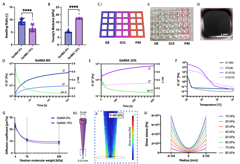 Figure 1. Characterization of bioinks
Computer simulations and experimental validations evaluated the flow distribution within the vascular channels and the diffusion of diluted species in the hydrogels. The results showed that within 10 minutes of perfusion initiation, the model could reach and maintain the target concentration, confirming that the perfusion system effectively ensures nutrient supply and oxygenation for the cells.
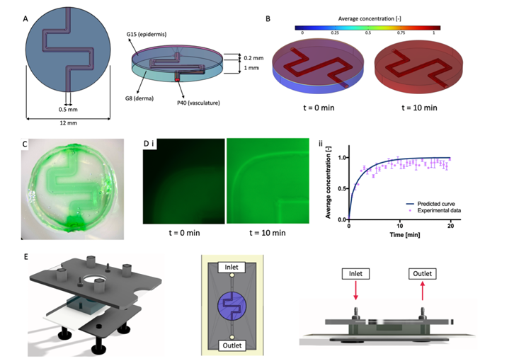 Figure 2. Model design and validation
The various components of the skin model exhibited good physiological functions. In the dermal layer, fibroblasts and endothelial cells co-cultured to form a dense microvascular network, with cell viability remaining above 90% after 21 days. The metabolic activity of the cells caused the Young’s modulus of G8 hydrogel to decrease from 8.65±0.81 kPa (without cells) to 3.1±0.2 kPa, and the extracellular matrix proteins secreted by fibroblasts, such as collagen (types I, III, IV), elastin, and fibronectin, significantly increased with culture time, indicating gradual tissue maturation.
In the epidermal layer, keratinocytes cultured in G15 reached a thickness of 246.6±22.8 μm after 21 days, occupying the entire printed volume. The expression of keratin 10 (CK10) significantly increased over time, indicating proper differentiation and stratification of keratinocytes. The transepithelial electrical resistance (TEER) values gradually increased over time, confirming the formation and maturation of the epithelial barrier. The vascular channels formed a complete endothelial barrier through co-culture of endothelial cells and fibroblasts, with vascular permeability significantly lower than the control group without endothelial cells after 21 days of perfusion, indicating good functional integrity of the endothelial barrier.
In wound healing studies, the model demonstrated excellent repair capabilities. Whether through damage caused by a 1mm biopsy punch penetrating the dermis and epidermis or linear scratch injuries on the epidermal surface, wound closure was achieved after 14 days of culture. Ki67 immunostaining showed active cell proliferation in the regenerated tissue, indicating that the model can simulate the repair processes of in vivo skin, including epithelial regeneration and dermal remodeling.
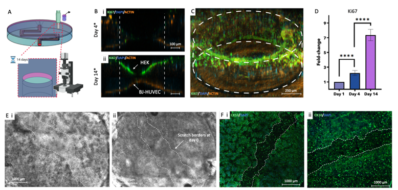 Figure 3. Evaluation of wound healing dynamics in the 3D vascularized skin model
The successful construction of this vascularized 3D bioprinted skin model provides a fully functional in vitro platform for skin tissue engineering research. It not only simulates the physiological structure and function of the skin but also reproduces key processes of wound healing, holding significant application value in cosmetics and drug development. The establishment of this model reduces reliance on animal experiments, providing a testing system that closely resembles the human physiological environment for evaluating product efficacy and safety, and is expected to promote advancements in the treatment of skin-related diseases and regenerative medicine. For researchers engaged in skin studies, pharmaceutical companies, and the general public concerned with skin health, this achievement undoubtedly brings new hope for skin health and injury repair.
References:
Maggiotto F, Dalla Valle E, Fietta A, Visentin LM, Giomo M, Cimetta E. 3D bioprinting of a perfusable skin-on-chip model suitable for drug testing and wound healing studies. Mater Today Bio. 2025;33:101974. Published 2025 Jun 13. doi:10.1016/j.mtbio.2025.101974
This article is for academic sharing only. Please indicate the source when reprinting. If there is any infringement, please contact WeChat: bioonSir for deletion or modification!
Click below 「Read the original text」, to visit the official website of Bio Valley for more biological-related information~