Understanding Pulmonary Valve Regurgitation

Understanding Pulmonary Valve Regurgitation

Pulmonary Valve Anatomy

Understanding Pulmonary Valve Regurgitation
The right ventricle is divided into inflow and outflow tracts, separated by the supraventricular crest. The supraventricular crest is a broad muscular ridge on the right ventricular wall, located between the right atrioventricular orifice and the pulmonary artery orifice. The outflow tract extends upward and narrows, resembling an inverted funnel, with smooth walls, known as the conus arteriosus. The upper end of the conus arteriosus is the opening leading to the pulmonary artery, referred to as the pulmonary artery orifice.

Anatomy of the Right Ventricular Outflow Tract to the Pulmonary Artery

Understanding Pulmonary Valve Regurgitation

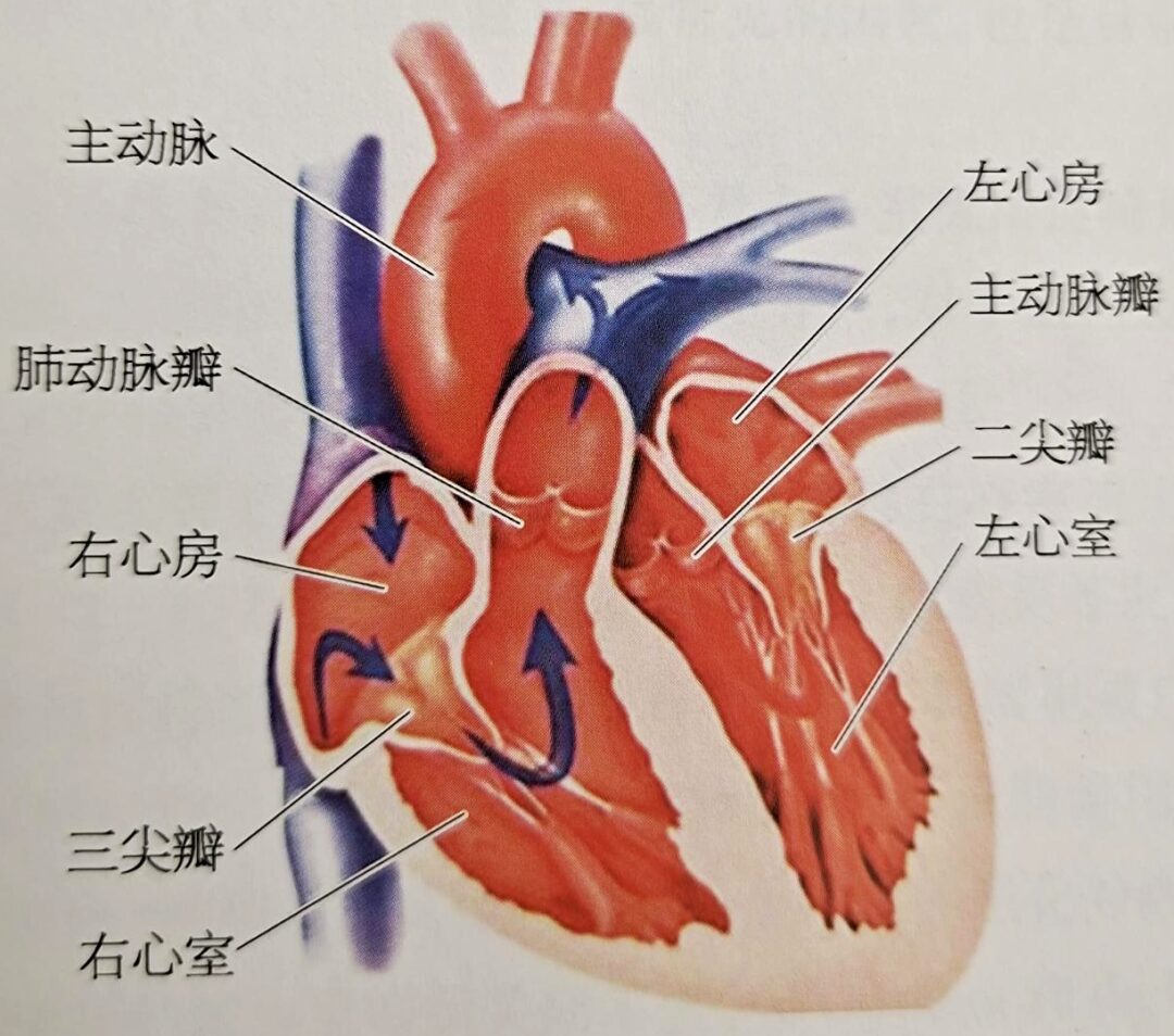
Surrounding the pulmonary artery orifice is the pulmonary valve annulus, which is attached to three crescent-shaped leaflets, known as the pulmonary valve. When the ventricle contracts, blood flows through the pulmonary valve into the pulmonary artery; during ventricular relaxation, the valve closes, preventing blood from flowing back into the ventricle. The three leaflets of the pulmonary valve, the cusps, and the annulus are relatively weak, and the annulus connects to the muscle of the right ventricular outflow tract (RVOT), without a direct fibrous continuity with the tricuspid valve. The three leaflets can be classified as the left, right, and anterior leaflets. The left leaflet is continuous with the septal band of the conus, while the right leaflet is continuous with the wall of the outflow tract. The inner half of the left and right leaflets is in contact with the aortic wall, and the junction between the left and right pulmonary valve leaflets corresponds to that of the aortic left and right leaflets, although these junctions do not connect at the same point, with the junction of the pulmonary valve being slightly higher. The anterior leaflet of the pulmonary valve is attached to the free wall of the right ventricle. Above the pulmonary valve is the pulmonary artery trunk. The pulmonary artery trunk is located within the pericardium and is a short, thick arterial trunk that originates from the right ventricular outflow tract, slanting left and posterior above the ascending aorta, and bifurcating into the left and right pulmonary arteries below the aortic arch.

Understanding Pulmonary Valve Regurgitation

What Are the Causes of Pulmonary Valve Regurgitation?

Understanding Pulmonary Valve Regurgitation
The causes of pulmonary valve regurgitation (PR) mainly include the following:

(1) Congenital pulmonary valve malformations:

This can include complete absence, unicuspid, bicuspid pulmonary valves, and other malformations, commonly seen in patients with Tetralogy of Fallot (TOF).

(2) Marfan syndrome:

This primarily affects the aortic and mitral valves, but reports indicate that about 26.9% of patients also involve the pulmonary valve, leading to pulmonary valve regurgitation.

(3) Cardiac tumors:

Papillary fibroelastoma is closely related to pulmonary valve disease, often presenting as multiple lobulated tumors attached to the endocardium via a stalk. This is usually seen on the ventricular side of the aortic or pulmonary valve. As the tumor size increases, it may cause moderate to severe pulmonary valve regurgitation, with or without pulmonary valve stenosis.

(4) Pulmonary valve annulus dilation:

Long-term pulmonary hypertension can lead to dilation of the pulmonary artery and annulus, causing pulmonary valve regurgitation. In patients with outflow tract obstruction, dilation may occur post-stenosis, leading to annulus enlargement and resulting in pulmonary regurgitation.

(5) Iatrogenic causes:

This is currently the most clinically significant and common factor, being the primary suitable population for PPVI (Percutaneous Pulmonary Valve Implantation).
Understanding Pulmonary Valve Regurgitation
The proportion of newborns with congenital heart disease combined with pulmonary valve annulus and right ventricular outflow tract malformations is nearly 20%. Common diseases include Tetralogy of Fallot, persistent arterial trunk, and pulmonary valve atresia. It has been previously suggested in foreign literature that surgical correction within one month of birth is crucial for the survival of these patients.
The 2018 AHA/ACC “Guidelines for Managing Adults with Congenital Heart Disease” recommend reconstructive procedures for the right ventricular outflow tract, including the use of a conduit with a valve and replacement with artificial biological valves.

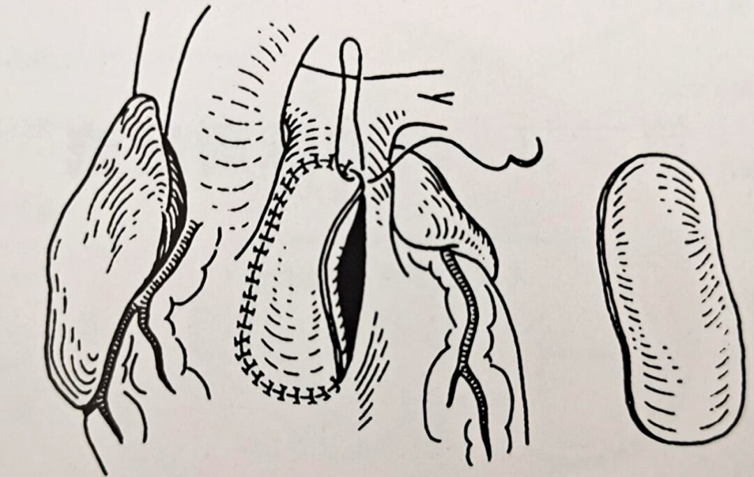
Patch for Transpulmonary Valve

Understanding Pulmonary Valve Regurgitation

In developed countries such as Europe and the United States, the proportion of conduit with a valve used previously is relatively high, while other techniques, including transvalvular patches, have also been widely used in the United States over the past decade. For patients receiving conduit implantation, although pulmonary regurgitation does not occur in the short term, over the long term (usually 5-15 years), the artificial vessel may calcify, leading to outflow tract obstruction, and the biological valve may degenerate, resulting in valve insufficiency or stenosis, or combined with the conduit may lead to aneurysmal dilation worsening valve insufficiency. Surveys show that in an average long-term follow-up of 20 years, mild to moderate pulmonary valve regurgitation can be present in almost all TOF patients, with a high incidence of severe pulmonary valve regurgitation, reaching 30%-40%.
In developing countries such as China, Brazil, and Argentina,reconstructive procedures for the right ventricular outflow tract primarily include simple pulmonary valve incision, closed balloon dilation of the pulmonary valve, and transvalvular patching, all of which may lead to pulmonary valve regurgitation. This is iatrogenic pulmonary valve regurgitation, with or without pulmonary valve stenosis. Among these, the most commonly used procedure istranspulmonary valve patching, which accounts for 60%-70%, and while alleviating pulmonary valve stenosis, it may lead to dilation of the pulmonary valve annulus, resulting in complete or partial loss of pulmonary valve function, which is the primary cause of iatrogenic pulmonary valve regurgitation.
In a study of 109 children after TOF surgery, long-term echocardiographic follow-up showed that the regurgitation rate after transvalvular patching reached 100%. In a cross-sectional study of 263 cases regarding pulmonary valve function after TOF surgery, it was found that only 8.4% of patients with mild pulmonary valve regurgitation, while the majority had moderate to severe pulmonary valve regurgitation. Additionally, simple pulmonary valve incision can also lead to varying degrees of damage to the leaflet structure, presenting as irregular thickening and the appearance of leaflet fenestration, severe rupture, and continuous damage to the junction, causing pulmonary valve regurgitation. Moreover, percutaneous balloon dilation can also lead to pulmonary valve regurgitation, but the degree of pulmonary valve regurgitation in these two types of surgeries is generally mild and clinically insignificant.
Transpulmonary Valve Patching is the most commonly used technique in China, which involves cutting the pulmonary valve annulus along the outflow tract axis, and then suturing a patch to widen it, which can lead to valve regurgitation while relieving stenosis.If the right leaflet of the pulmonary valve can be preserved by cutting along the left leaflet-anterior leaflet junction, it may reduce the degree of postoperative pulmonary valve regurgitation.
Understanding Pulmonary Valve Regurgitation

Pulmonary Valve Regurgitation Pathophysiology

Understanding Pulmonary Valve Regurgitation
Simple pulmonary valve regurgitation is generally well tolerated in the early stages. Studies show that among patients with congenital pulmonary malformations causing valve insufficiency, only 6% develop symptoms within 20 years, and 49% develop symptoms within 40 years. This study has been cited by later scholars as the basis for using transvalvular patching in RVOT enlargement surgery for TOF. Patients with pulmonary valve regurgitation after TOF surgery often do not present symptoms in the first 10-20 years.However, chronic pulmonary valve regurgitation can lead to increased right heart load, right heart enlargement, subsequently causing right heart failure, arrhythmias (ventricular premature contractions, ventricular tachycardia, ventricular fibrillation, atrial flutter, atrial fibrillation), and even sudden death. Additionally, due to the enlarged right heart compressing the left heart, it can lead to left heart dysfunction (decreased left ventricular ejection fraction, reduced exercise tolerance), further worsening the patient’s clinical status.Moreover, some TOF postoperative patients may experience aneurysmal dilation of the right ventricular outflow tract due to the patch’s lack of contractile function, further exacerbating right heart dysfunction. Sudden cardiac death is the most severe adverse event in patients with pulmonary valve regurgitation, with an incidence of 0.06% per year within 10 years after TOF repair, but it can reach 0.20% per year after 10 years.Risk factors include RVOT patching, male gender, high NYHA classification, and QRS duration >180ms.Malignant ventricular arrhythmias are the primary cause of sudden death in these patients.
A study involving 793 TOF patients showed that after an average follow-up of 21 years, 33 patients had recorded sustained ventricular tachycardia, 29 had atrial fibrillation or flutter, and 16 patients experienced sudden cardiac death. Patients after TOF repair with RVOT aneurysmal dilation, tricuspid regurgitation, or pulmonary valve regurgitation are more likely to develop sustained ventricular tachycardia. Studies indicate that the QRS duration in patients after TOF repair is positively correlated with right ventricular size, with significantly enlarged right ventricles and QRS duration >180ms being risk factors for sustained ventricular tachycardia. It should also be noted that the pathophysiological changes in patients with pulmonary hypertension combined with pulmonary valve regurgitation are primarily caused by pulmonary hypertension rather than by pulmonary valve regurgitation, and treatment should focus on correcting pulmonary hypertension.
Understanding Pulmonary Valve Regurgitation

Choosing the Timing of Intervention

Understanding Pulmonary Valve Regurgitation
Due to the long-term adverse effects of pulmonary valve regurgitation on patient prognosis, the treatment method of pulmonary valve replacement (PVR) has been proposed, including surgical PVR and PPVI.
(1) PVR
Regarding the long-term prognosis of surgical PVR, a meta-analysis including 48 studies with 3118 surgical PVR patients showed that the 30-day postoperative mortality rate was 0.87%, the 5-year mortality rate was 2.20%, and the rate of reoperation for pulmonary valve replacement within 5 years was 4.90%. The benefits of surgical PVR includereduced right ventricular volume, improved right and left heart function, shortened QRS duration, and symptom improvement. Thus, surgical PVR indeed provides significant benefits to patients with pulmonary valve regurgitation after TOF surgery.
Understanding Pulmonary Valve Regurgitation
Timing of Surgery: There is an upper limit to the timing of surgery. Therrien et al. first proposed the concept of a “surgical upper threshold”. Their research found that in patients with pulmonary valve regurgitation, preoperative right ventricular end-diastolic volume index (RVEDVI) >170ml/m² or right ventricular end-systolic volume index (RVEDSI) >85ml/m², the right ventricular size cannot be reduced to normal (RVEDVI <100ml/m²) postoperatively.
Patients with preoperative RVEDVI <162ml/m² or RVEDSI <82ml/m² can have their right ventricular size reduced to normal after surgical PVR. Geva found that using a preoperative RVEDSI <90ml/m² as a threshold yields the best improvement in right ventricular size and function post-surgery. Studies indicate that the optimal upper threshold for PVR surgery is RVEDVI <163ml/m² or RVEDSI <80ml/m². These findings suggest that patients with pulmonary valve regurgitation should undergo surgery before the right ventricle enlarges to these upper threshold levels; otherwise, the surgical outcomes will be poor.
The 2008 AHA/ACC “Guidelines for the Management of Adults with Congenital Heart Disease” and the 2014 AHA/ACC “Guidelines for the Management of Valvular Heart Disease” recommend that indications for PVR include:
① Severe pulmonary valve regurgitation with symptoms or decreased exercise tolerance (Class I indication, Level B evidence);
② Asymptomatic severe pulmonary valve regurgitation with any of the following: moderate to severe right ventricular enlargement or dysfunction, moderate to severe tricuspid regurgitation, persistent atrial or ventricular arrhythmias (Class IIa indication, Level B and C evidence).
Indications for PVR in the 2010 European Society of Cardiology guidelines are:
① Severe pulmonary valve regurgitation or stenosis with symptoms (Class I indication, Level C evidence);
② Asymptomatic severe pulmonary valve regurgitation or stenosis with any of the following: decreased exercise tolerance, progressive right ventricular enlargement or dysfunction, progressive tricuspid regurgitation, persistent atrial or ventricular arrhythmias (Class IIa indication, Level C evidence).
In these guidelines, there is no clear definition for moderate to severe or progressive right ventricular enlargement or dysfunction, which may be related to the current lack of clinical evidence. However, in the 2014 consensus of multiple American societies, the indications for transcatheter valve repair and replacement procedures clearly state that PVR indications include symptomatic severe pulmonary valve regurgitation or asymptomatic patients with cardiac MRI showing RVEDVI >150ml/m², pulmonary regurgitation index >40%, or ventricular ejection fraction <40%.
(2) PPVI
Compared to surgical procedures, PPVI is less invasive and carries lower surgical risks. Currently, the advantages of PPVI include:
① Improvement in heart function and symptoms, enhancing quality of life;
② Potential reduction in sudden death risk for some patients, thereby improving prognosis;
③ Delay in the timing of surgical procedures, reducing the number of surgeries required for patients, particularly suitable for high-risk surgical patients.
If patients experience valve dysfunction after PPVI, they can undergo PPVI again; if re-PPVI is not possible, surgical intervention can be performed. Thus, PPVI can delay the timing of surgical procedures and reduce the number of surgical PVRs, making it a more attractive treatment option.
Understanding Pulmonary Valve Regurgitation
In summary, based on domestic clinical practice experience and relevant literature, the first edition of the “Expert Recommendations for Percutaneous Pulmonary Valve Implantation in China” published in 2016 preliminarily summarizes the indications and contraindications for PPVI applicable to the Chinese population.

1. Indications

(1) Moderate to severe pulmonary valve regurgitation following surgical correction of congenital heart disease with RVOT stenosis.

(2) Patients with symptoms related to right ventricular outflow tract stenosis, including decreased exercise tolerance or right heart failure; or asymptomatic patients with any of the following: ① Moderate or greater functional tricuspid regurgitation; ② Cardiac MRI showing RVEDVI ≥130ml/m²; ③ Cardiac MRI showing right ventricular ejection fraction <45%, QRS duration ≥160ms; ④ Persistent atrial or ventricular arrhythmias.

(3) Anatomically suitable for PPVI.

(4) Age ≥10 years or weight ≥25kg.

2. Contraindications

(1) Pulmonary hypertension [mean pressure ≥25mmHg (1mmHg=0.133kPa)].

(2) Severe pulmonary artery (PA) or branch stenosis.

(3) Anatomical assessment unsuitable, including inability to deliver the valve through vascular access or inability to place the valve in the RVOT-PA, or preoperative evaluation suggests that the valve stent may compress the coronary artery.

(4) Presence of contraindications for catheterization.

Understanding Pulmonary Valve Regurgitation

END

Source: Long Yuliang, Zhang Xiaochun, Pan Wenzhi. “Transcatheter Heart Valve Procedures”. Shanghai Scientific and Technical Publishers

References

1.Shimazaki Y, Blackaone EH, Kirklin JW. The natural history of isolated congenital pulmonary valve incompetence: surgical, implications[J]. Thorac Cardiovase Surg, 1984, 32:257-259.

2.McElhinney DB. Hennesen JT. The Melody valve and Ensemble delivery system for transcatheter pulmonary valve replacement[J]. Ann NY Acad Sci, 2013, 1291:77-85.

3.Murphy JG. Gersh BJ, Mair DD, et al. Long-term outcome in patients undergoing surgical repair of tetralogy of Fallot[J]. N Engl J Med, 1993, 329:593-599.

4.Warnes CA, Williams RG, Bashore TM, et al. ACC/AHA 2008 guidelines for the management of adults with congenital heart disease: a report of the American College of Candiology/American Heart Association Task Force on Practice Guidelines [J]. J Am Coll Cardiol, 2008, 52:e143-263.

5.d’Udeiem Y, Ovaert C, Grandjen F, et al. Tetralogy of Fallot: transannular and right ventricular patching equally affect late functional status[J]. Circulation, 2000, 102(19 Suppl 3):Ⅲ116-Ⅲ122.

6.de Ruijter FT, Weenink I, Hitchcock FJ, et al. Right ventricular dysfunction and pulmonary valve replacement after correction of tetralogy of Fallot[J]. Ann Thorac Surg, 2002, 73(6):1794-1800.

7.Hui Wei, Sun Bin, Huang Guoying, et al. Evaluation of the mid- and long-term efficacy of 109 cases of tetralogy of Fallot after surgery[J], Chinese Journal of Ultrasound Medicine, 2001 (4): 33-35.

8.Jiang Rui, Yan Jun, Li Shoujun, et al. Clinical analysis of 178 cases of tetralogy of Fallot repair[J]. Clinical Journal of Cardiovascular Diseases, 2011 (9): 702-704.

9.Ji Guangyu, Xu Zhiyun, Wang Zhongnong, et al. Long-term efficacy of surgical treatment for adult tetralogy of Fallot[J]. Chinese Journal of Thoracic and Cardiovascular Surgery, 2010 (4): 287-291.

10.Xie Zhaofeng, Zhang Zhiwei, Xu Yanmei, et al. Long-term efficacy and complications after repair of tetralogy of Fallot[J]. Clinical Journal of Cardiovascular Diseases, 2013, 29 (8): 618-620.

11.Long Yuliang, Pan Wenzhi, Zhan Zhi, et al. Analysis of pulmonary valve function in patients after surgical repair of tetralogy of Fallot[J]. Chinese Journal of Cardiovascular Diseases, 2017, 45 (8): 722-725.

12.Chaturvedi Re, Redington AN. Pulmonary regurgitation in congenital heart disease[J]. Heart, 2007, 93:880-889.

13.Nollert GD, Dabritz SH, Schmoeckel M, et al. Risk factors for sudden death after repair of tetralogy of Fallot[J]. Ann Thorac Surg, 2003, 76:1901-1905.

14.Gatzoulis MA, Balaji S, Webber SA, et al. Risk factors for arrhythmia and sudden cardiac death late after repair of tetralogy of Fallot: a multicentre study[J]. Lancet, 2000, 356:975-91.

15.Harrison DA. Harris L. Siu Sc. at al. Sustained ventricular in adult patients late after repair of tetralogy of Fallot[J]. J Am Coll Cardiol, 1997, 30:1368-1373.

16.Gatzoulis MA. Till JA, Somerville J, et al. Mechanoelectrical interaction in tetralogy of Fallot. QRS prolongation relates to right ventricular size and predicts malignant ventricular arrhythmias and sudden death[J]. Circulation, 1995, 92:231-237.

17. Ferraz Caralcanti PE, Sa MP, Santos CA, et al. Pulmonary valve replacement after operative repair of tetralogy of Fallot: meta-analysis and meta-regression of 3,118 patients from 48 studies[J]. Journal of the American College of Cardiology, 2013, 62(23):2227-2243.

Understanding Pulmonary Valve Regurgitation

● Observational Analysis of Echocardiography in Patients with Atrial Fibrillation

● The Relationship Between the Left Atrial Appendage and Atrial Fibrillation

● What Is Mitral Regurgitation? You Will Understand After Reading!

● Understanding the Mitral Valve!

Understanding Pulmonary Valve Regurgitation

Leave a Comment