The Medical Imaging Center of Shaoyang University Affiliated First Hospital currently owns one Siemens 1.5T Magnetic Resonance Imaging (MRI) machine, one Canon 80-slice 160-layer spiral CT, one Siemens 64-slice 128-layer spiral CT, and one GE 16-slice spiral CT. Additionally, there are two Philips flat-panel digital subtraction angiography (DSA) machines (one yet to be installed), one United Imaging dual flat-panel DR, one Philips flat-panel DR, one Canon multifunctional digital gastrointestinal machine (RF), one digital panoramic dental machine, one Hologic digital mammography machine, and one GE bone densitometer. These machines provide high-resolution, high-definition imaging examinations for various organs and systems of the body, enabling MRA, MRCP, CTA, CT coronary imaging, three-dimensional imaging of various organs, and various interventional diagnostic and therapeutic procedures. They provide essential imaging data for the diagnosis of diseases and the selection of treatment methods.
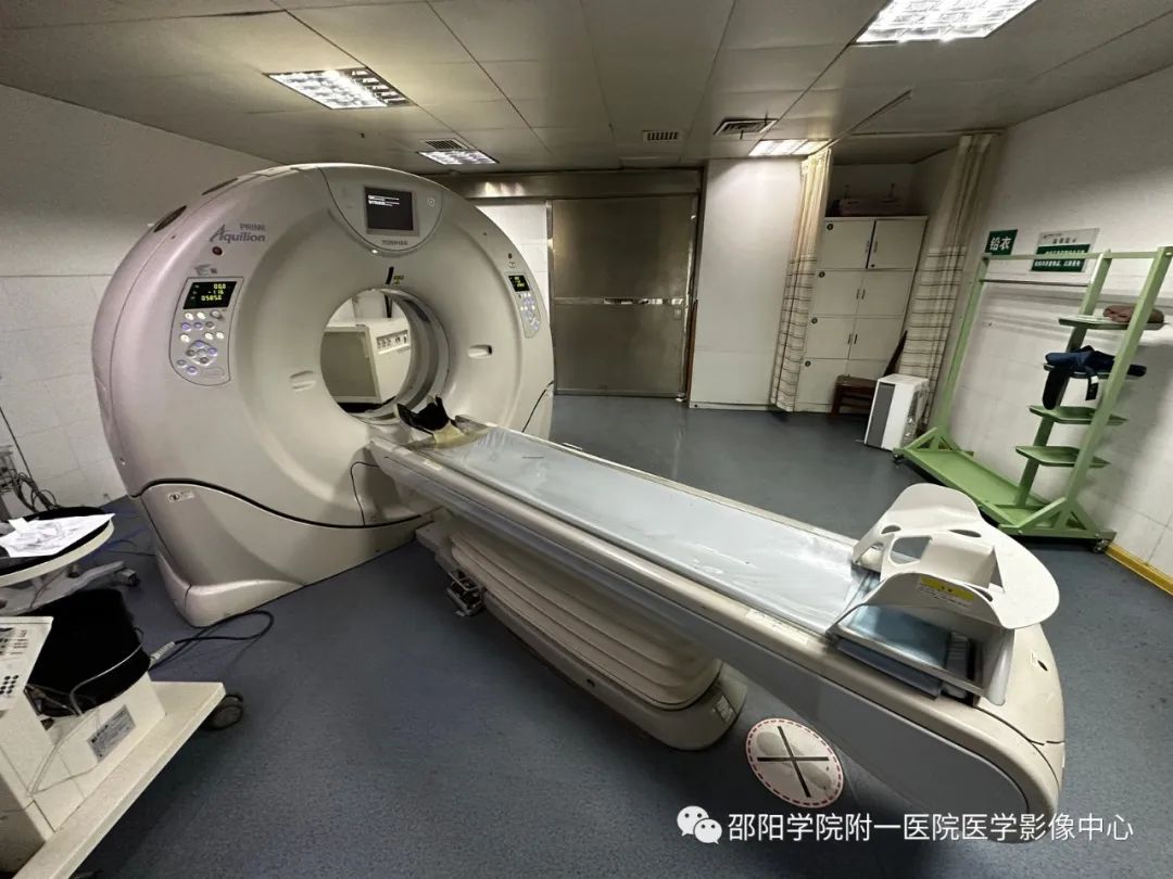 Canon 80-slice 160-layer spiral CT
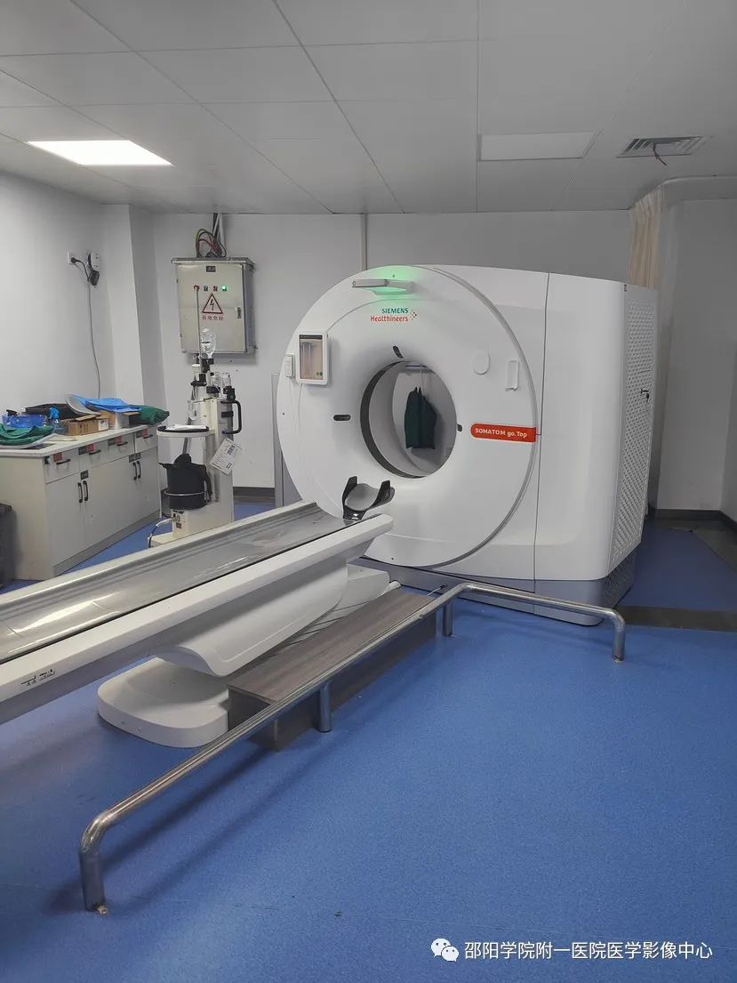
Siemens 64-slice 128-layer spiral CT
CT:Indications: Can perform routine CT scans and enhanced scans of conventional sites, and has capabilities for whole-body cardiovascular imaging (CTA) and urinary system imaging (CTU), useful for lung nodule analysis and early lung cancer screening. It can achieve maximum intensity projection (MIP), three-dimensional imaging of multiple body parts (VR), virtual endoscopy (VE), and multi-planar reconstruction (MPR) among various post-processing imaging techniques. Advantages: clearer, safer, more sensitive, and broader range imaging.
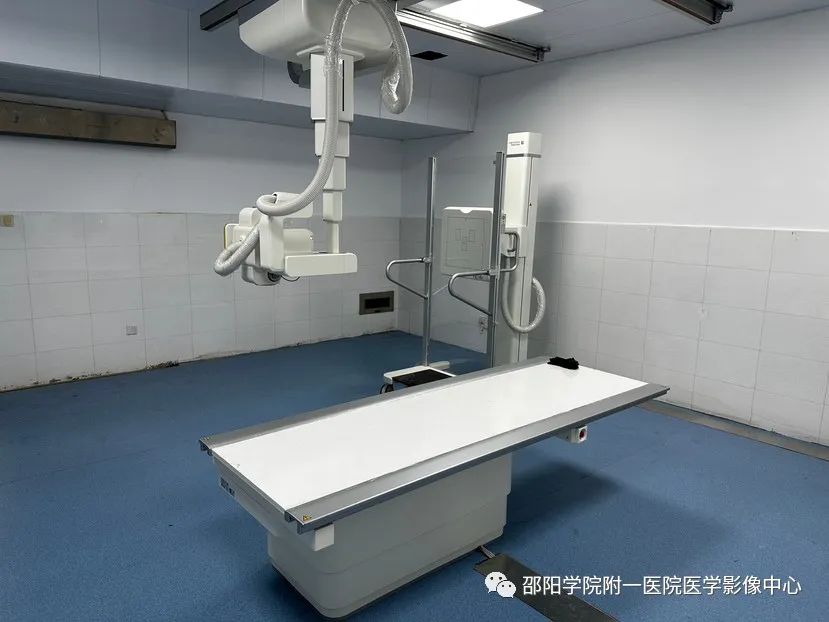
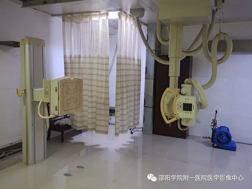
United Imaging dual flat-panel DR
DR:Used for routine imaging of various body parts. The images are clear, detailed, and high in contrast, minimizing the chances of missing lesions; X-ray radiation is reduced, lessening the harm of ionizing radiation to the human body; imaging speed is fast, and the image post-processing capabilities are powerful, allowing doctors to extract rich and reliable clinical diagnostic information based on the patient’s specific condition, providing a good diagnostic basis for the diagnosis of diseases, especially for the early detection of lesions.
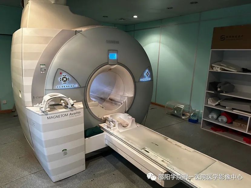
Siemens 1.5T superconducting MRI
MRI:Indications: For lesions of the brain, eyes, nose, throat, spine, joints, lungs, mediastinum, upper abdomen and retroperitoneal lesions, pelvic lesions, soft tissue lesions, vascular lesions, etc. Particularly unique in examining the brain, spinal cord, bone and joint soft tissues, and internal organs. Advantages: high resolution, sensitive display of lesions, non-invasive, painless, and no radiation exposure.
Canon 80-slice 160-layer spiral CT
Siemens 64-slice 128-layer spiral CT
CT:Indications: Can perform routine CT scans and enhanced scans of conventional sites, and has capabilities for whole-body cardiovascular imaging (CTA) and urinary system imaging (CTU), useful for lung nodule analysis and early lung cancer screening. It can achieve maximum intensity projection (MIP), three-dimensional imaging of multiple body parts (VR), virtual endoscopy (VE), and multi-planar reconstruction (MPR) among various post-processing imaging techniques. Advantages: clearer, safer, more sensitive, and broader range imaging.
United Imaging dual flat-panel DR
DR:Used for routine imaging of various body parts. The images are clear, detailed, and high in contrast, minimizing the chances of missing lesions; X-ray radiation is reduced, lessening the harm of ionizing radiation to the human body; imaging speed is fast, and the image post-processing capabilities are powerful, allowing doctors to extract rich and reliable clinical diagnostic information based on the patient’s specific condition, providing a good diagnostic basis for the diagnosis of diseases, especially for the early detection of lesions.
Siemens 1.5T superconducting MRI
MRI:Indications: For lesions of the brain, eyes, nose, throat, spine, joints, lungs, mediastinum, upper abdomen and retroperitoneal lesions, pelvic lesions, soft tissue lesions, vascular lesions, etc. Particularly unique in examining the brain, spinal cord, bone and joint soft tissues, and internal organs. Advantages: high resolution, sensitive display of lesions, non-invasive, painless, and no radiation exposure.
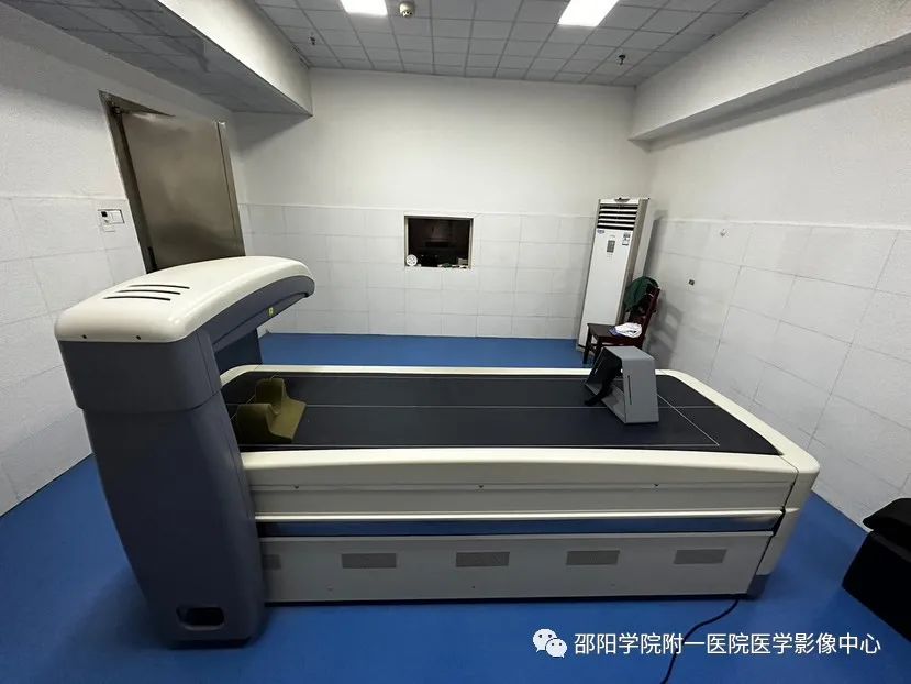 Bone Density: It accurately evaluates the health status of the entire skeletal system by measuring the bone mass of the left forearm (right side for left-handed individuals), and is a commonly used method for the early diagnosis of osteoporosis, disease monitoring, and evaluation of drug efficacy. It facilitates clinical medication guidance and targeted treatment and prevention for the subjects. This instrument has advantages such as fast scanning speed, high precision and accuracy, and low radiation dose, and has been widely used in clinical practice, recognized as the “gold standard” for bone density testing.
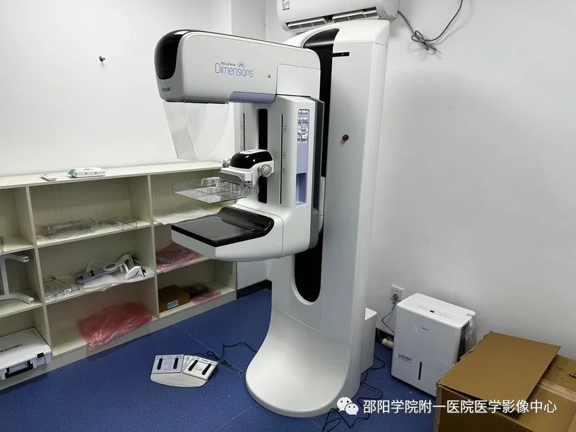
Digital mammography machine
Digital mammography: Mammography is a low-dose X-ray photography examination that is simple, safe, reliable, and has high resolution and good repeatability, not limited by age or body shape, and is currently a routine examination for breast checks. It can detect certain precancerous lesions and perform follow-up imaging for dynamic observation; it can detect breast cancer early, even identifying clinically undetectable hidden breast cancers; it can also reliably distinguish between benign breast lesions and malignant tumors. It is typically recommended to perform the examination 3-7 days after the menstrual period.
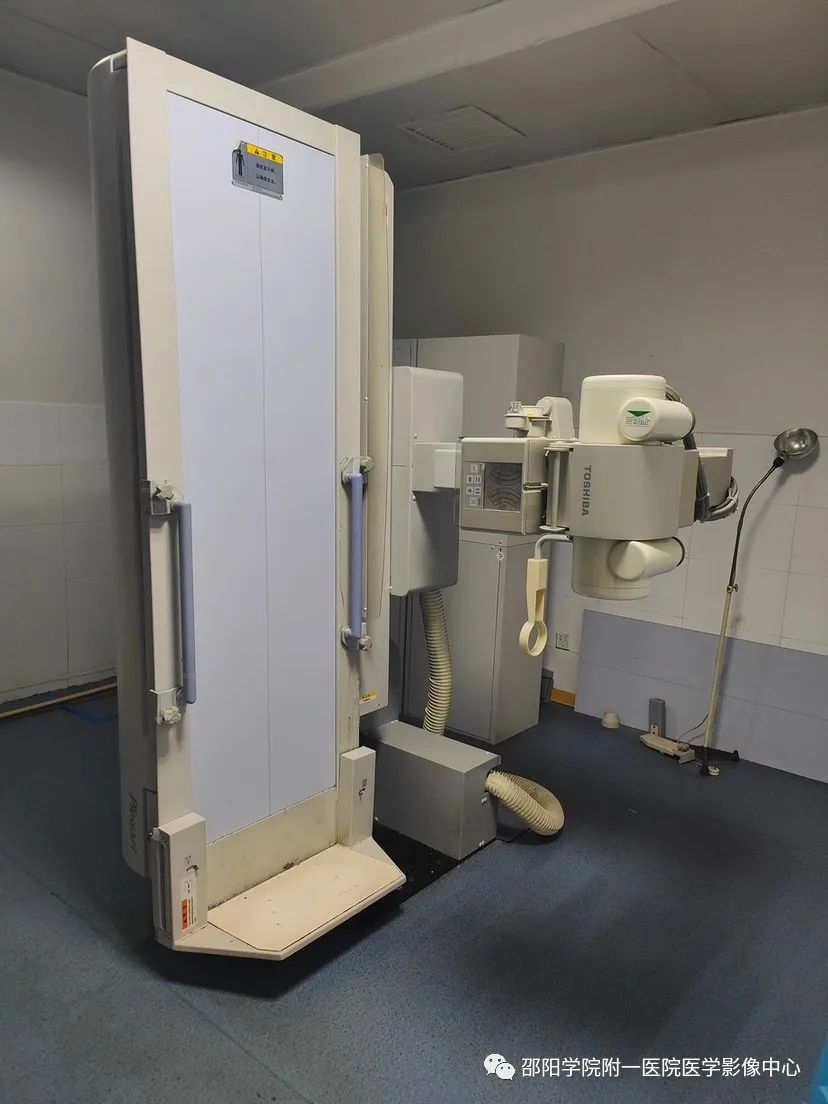
Digital gastrointestinal X-ray machine
Digital gastrointestinal: Used for routine chest and abdominal fluoroscopy, esophagus, upper digestive tract, and complete digestive tract barium meal examinations, barium enema examinations, “T” tube and fistula angiography examinations, urinary system retrograde angiography, and uterine tube angiography among various contrast examinations. This machine has a high-frequency generator, ball tube, and integrated digital imaging system, which features good image quality, digital image acquisition, and clearer images after digital processing, allowing doctors to observe subtle lesions under various conditions, thus improving accuracy.
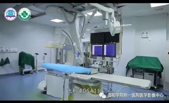
Philips flat-panel DSA machine
DSA: Indications: (1) Interventional treatment of heart disease: such as coronary angiography, stent implantation, temporary pacemaker implantation, permanent pacemaker implantation, and radiofrequency ablation treatment for rapid arrhythmias. (2) Interventional treatment of tumors: embolization treatment for liver cancer, lung cancer, stomach cancer, and stent placement for esophageal cancer with stenosis. (3) Interventional treatment of gynecological diseases: embolization for postpartum hemorrhage, tubal recanalization, and embolization treatment for uterine fibroids. (4) Interventional treatment of peripheral vascular diseases: emergency embolization for various massive hemorrhages, stent implantation for vascular tumors, and interventional treatment for vasculitis and vascular malformations. Advantages: clear imaging, stable performance, low radiation. The interventional procedures are simple, safe, effective, minimally invasive, and with fewer complications.
Bone Density: It accurately evaluates the health status of the entire skeletal system by measuring the bone mass of the left forearm (right side for left-handed individuals), and is a commonly used method for the early diagnosis of osteoporosis, disease monitoring, and evaluation of drug efficacy. It facilitates clinical medication guidance and targeted treatment and prevention for the subjects. This instrument has advantages such as fast scanning speed, high precision and accuracy, and low radiation dose, and has been widely used in clinical practice, recognized as the “gold standard” for bone density testing.
Digital mammography machine
Digital mammography: Mammography is a low-dose X-ray photography examination that is simple, safe, reliable, and has high resolution and good repeatability, not limited by age or body shape, and is currently a routine examination for breast checks. It can detect certain precancerous lesions and perform follow-up imaging for dynamic observation; it can detect breast cancer early, even identifying clinically undetectable hidden breast cancers; it can also reliably distinguish between benign breast lesions and malignant tumors. It is typically recommended to perform the examination 3-7 days after the menstrual period.
Digital gastrointestinal X-ray machine
Digital gastrointestinal: Used for routine chest and abdominal fluoroscopy, esophagus, upper digestive tract, and complete digestive tract barium meal examinations, barium enema examinations, “T” tube and fistula angiography examinations, urinary system retrograde angiography, and uterine tube angiography among various contrast examinations. This machine has a high-frequency generator, ball tube, and integrated digital imaging system, which features good image quality, digital image acquisition, and clearer images after digital processing, allowing doctors to observe subtle lesions under various conditions, thus improving accuracy.
Philips flat-panel DSA machine
DSA: Indications: (1) Interventional treatment of heart disease: such as coronary angiography, stent implantation, temporary pacemaker implantation, permanent pacemaker implantation, and radiofrequency ablation treatment for rapid arrhythmias. (2) Interventional treatment of tumors: embolization treatment for liver cancer, lung cancer, stomach cancer, and stent placement for esophageal cancer with stenosis. (3) Interventional treatment of gynecological diseases: embolization for postpartum hemorrhage, tubal recanalization, and embolization treatment for uterine fibroids. (4) Interventional treatment of peripheral vascular diseases: emergency embolization for various massive hemorrhages, stent implantation for vascular tumors, and interventional treatment for vasculitis and vascular malformations. Advantages: clear imaging, stable performance, low radiation. The interventional procedures are simple, safe, effective, minimally invasive, and with fewer complications.
Images: Department equipment photos
Text: Xiao Jiarong, Zeng Ying
Editor: Guo Huanhui
Reviewers: Tang Yongjun, Fan Jinhua, Guo Jianwei, Liu Qianjin
Planning: Shaoyang University Affiliated First Hospital Medical Imaging Department