
01
Supporting New Research Discoveries
Nanoinsights
The nucleolus, as the largest non-membrane organelle within the nucleus, serves as the core factory for ribosomal RNA synthesis and processing. Its multi-level structure (fibrillar center FC / dense fibrillar component DFC / peripheral dense fibrillar component PDFC / granular component GC) has long been believed to be closely related to the maturation processes of the small subunit (SSU) and large subunit (LSU) of ribosomes.
Scientific Question
The spatiotemporal functional correlation between pre-rRNA (precursor ribosomal RNA) processing and nucleolar substructures in adapting to changes in cellular physiological states (such as alterations in proliferation rates) remains a key scientific question that has not been fully resolved.
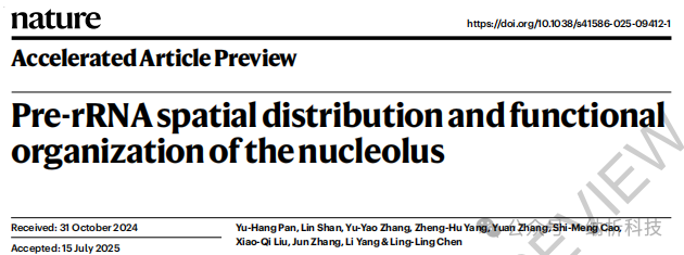
In July 2025,the research team led by Chen Lingling at the Center for Excellence in Molecular Cell Science, Chinese Academy of Sciences, published a paper titled “Pre-rRNA spatial distribution and functional organization of the nucleolus” in Nature.The research team revealed for the first time:
1️⃣Mechanism of compartmentalized processing: The processing of the small subunit (SSU) primarily occurs in the FC/DFC/PDFC regions, while the processing of the large subunit (LSU) mainly takes place during the transport from PDFC to GC, overturning the traditional “uniform diffusion” model;
2️⃣The pivotal role of the 5′ ETS: In slowly proliferating cells, the obstruction of 5′ ETS processing directly leads to the reorganization of the FC/DFC structure, proving that SSU processing centered around 5′ ETS is crucial for maintaining nucleolar structural stability;
3️⃣Evidence of evolutionary advantage: Compared to the bipartite nucleolus of amniote animals like zebrafish (where FC/DFC fuse into a fibrillar zone FZ), the multi-level nucleolus in humans enhances pre-rRNA processing efficiency by seven times through the establishment of nested FC/DFC structures, providing an evolutionary advantage for ribosome biogenesis.
02
Contribution of Multi-SIM in this Article
Nanoinsights
Experiment 1
To explore the impact of cellular proliferation status on the transport rate of newly synthesized pre-rRNA within the nucleolus, researchers conducted a 5-EU pulse labeling on rapidly proliferating cells (H9 human pluripotent stem cells) and slowly proliferating cells (SH-SY5Y neuroblastoma cells, D30 differentiated arcuate neurons) to track the spatial distribution changes of pre-rRNA within 0-60 minutes.
Conclusion 1
Through Multi-SIM imaging, it was found that:
1
Rapidly proliferating cells (H9)
pre-rRNA rapidly transferred from FC/DFC to GC within 30 minutes;
2
Slowly proliferating/differentiated cells
The transport rate of pre-rRNA significantly decreased (2-fold reduction in SH-SY5Y, 5-fold reduction in D30 neurons), with signals retained in the DFC-PDFC region.
This indicates that a decrease in cellular proliferation rate leads to obstruction of newly synthesized pre-rRNA outflow. Combined with other experimental results, it is evident that SSU processing centered around 5′ ETS is crucial for the correct assembly of FC/DFC.
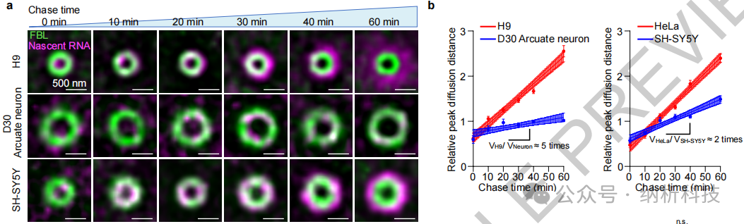
Figure Caption: a. Representative average SIM images of newly synthesized pre-rRNA and FBL in H9, SH-SY5Y, and D30 arcuate neurons during the tracking time (0-60 min). Scale bar: 500 nm. b. Statistical graph showing the change in peak diffusion distance of newly synthesized pre-rRNA relative to FBL over time in HeLa, SH-SY5Y, H9, and D30 arcuate neurons.
Experiment 2
To verify the necessity of 5′ ETS processing for maintaining the FC/DFC structure of the nucleolus, antisense oligonucleotides (ASOs) were used to target and interfere with the 5′ ETS cleavage sites (A0 and 1 sites) of SSU pre-rRNA, disrupting the FC/DFC structure. ASOs targeting LSU processing sites (ASO-U8, ASO-3′ ETS) and scrambled ASO were used as controls to observe changes in the nucleolar structure of HeLa cells.
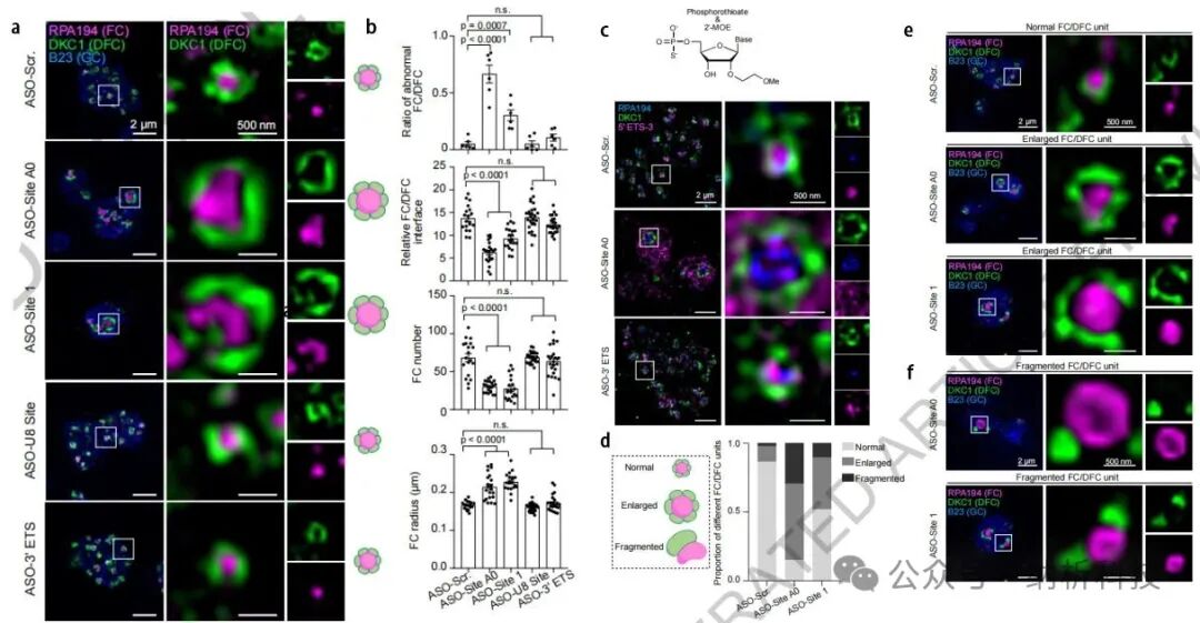
Figure Caption: a. Representative SIM images of HeLa cells treated with ASOs, indicating FC (RPA194, magenta), DFC (DKC1, green), and GC (B23, blue). b. Statistical results of FC/DFC reorganization after ASO treatment. e. Further displays SIM fluorescence imaging of three different morphologies of FC/DFC after ASO treatment: normal, enlarged, and fragmented; d. Schematic diagram and proportions of different morphologies under various treatment conditions. c. Fluorescent localization images of antisense oligonucleotides (ASO) containing thio-phosphoramidate and Z-MOE modifications and 5′ ETS-3 (magenta) in HeLa cells treated with ASOs.
Conclusion 2
Multi-SIM fluorescence imaging results show:
01
Abnormal FC/DFC Morphology
The nucleolus exhibited two main abnormal morphologies: “Enlarged” and “Fragmented”.
★ Normal: Compact FC/DFC nested structure.
★ Enlarged: Expansion of FC volume, thickening of DFC enveloping layer.
★ Fragmented: FC/DFC breaking into discrete points.
Among them, ASO-Site A0 had a more significant impact, with approximately 50% of nucleoli exhibiting an Enlarged morphology and 20% a Fragmented morphology. Inhibition of SSU pre-rRNA processing leads to a reduction in the relative contact interface of FC/DFC, a decrease in the number of FCs, and an increase in volume. (Figures a, b, e, d)
02
Abnormal pre-rRNA Localization
The 5′ ETS-3 signal diffused and moved outward to the outer layer of DFC (Figure c), indicating that obstruction of SSU processing leads to abnormal localization of pre-rRNA.
03
Summary of Research Findings
Nanoinsights
This research not only reveals the precise operational mechanisms of the nucleolus—regional division of labor in pre-rRNA processing, functional interactions between 5′ ETS processing and nucleolar structure, and the evolutionary advantages of multi-level nucleoli—but also reshapes our understanding of the spatial assembly of the nucleolus,highlighting the irreplaceable role of Multi-SIM in the dynamic analysis of organelles: its ultra-high resolution (~85 nm) and low phototoxicity live-cell imaging capabilities make it a powerful tool for deciphering the spatiotemporal codes of subcellular compartments.In the future, with continuous upgrades in imaging technology, we may unlock more operational codes of cellular “microfactories” and unveil finer regulatory mechanisms of life activities.



Nanoinsights Technology|Global leader in super-resolution microscopy technology
Recommended Reading
Nanoinsights Technology Shines at the 21st Biophysical Conference
Nanoinsights Technology Leads the Domestic SIM Super-Resolution Market, Showcasing China’s Manufacturing Strength
Research Insights|Multi-SIM Supports Research Directions in Liquid-Liquid Phase Separation (Part 1)