


The team led by Hu Bing from the Department of Gastroenterology, West China Hospital, Sichuan University, published an article titled “Effect of an artificial intelligence-assisted system on endoscopic diagnosis of superficial oesophageal squamous cell carcinoma and precancerous lesions: a multicentre, tandem, double-blind, randomised controlled trial” in The Lancet Gastroenterology & Hepatology on November 10, 2023 (scan the QR code to read the original text). This study reveals that the AI (artificial intelligence) system developed by the team can significantly improve the detection rate of early esophageal squamous cell carcinoma, has the potential to reduce the risk of missed lesions, and demonstrates good safety. This study is the first report in the world to evaluate the real-time diagnostic efficacy of AI systems in assisting early esophageal squamous cell carcinoma diagnosis in real clinical practice, and it currently includes the largest number of cases for AI-assisted diagnosis of early esophageal squamous cell carcinoma internationally.
Esophageal squamous cell carcinoma is a malignant tumor of the digestive tract that threatens the health of residents in our country. The 5-year survival rate of patients with early esophageal squamous cell carcinoma after minimally invasive treatment can reach over 90%, while the overall 5-year survival rate of advanced patients is less than 20%. Therefore, improving the early diagnosis rate of esophageal squamous cell carcinoma is crucial for improving patient prognosis. Gastrointestinal endoscopic screening is an important means for diagnosing early esophageal squamous cell carcinoma. Although the screening for early esophageal squamous cell carcinoma is relatively widespread at various levels of hospitals, there are significant differences in regional medical levels, and there is a severe shortage of experienced endoscopists, which poses a severe challenge for most grassroots hospitals in screening for early esophageal squamous cell carcinoma, raising concerns about missed diagnoses. In recent years, AI has developed rapidly in the field of medical auxiliary diagnosis, achieving significant progress in assisting endoscopic diagnosis of early esophageal squamous cell carcinoma.
The team led by Professor Hu Bing has been committed to the applied basic research of AI-assisted diagnosis of early esophageal squamous cell carcinoma for the past five years. By collaborating with universities and excellent enterprises, they have established an “industry-academia-research-medical” system, creating a multifunctional AI system for real-time assistance in diagnosing early esophageal squamous cell carcinoma that can determine lesion properties, outline lesion boundaries, and assess lesion infiltration depth. Unlike similar products reported domestically and internationally that require an external display, this system is directly “embedded” in the endoscope display, allowing for single-screen use and flexible signal source switching without changing the operating habits of endoscopists. During gastroscopy, the AI system can identify early esophageal squamous cell carcinoma in real-time, marking suspected lesion areas in polygonal form and displaying the probability value of the suspected lesion in the upper left corner of the endoscopic display (Figure 1), thus assisting doctors in diagnosing early esophageal squamous cell carcinoma in real-time.
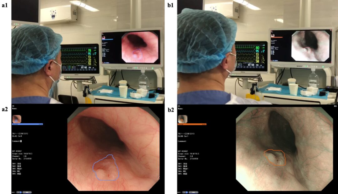
Figure 1: Example of AI System Assisting Doctors in Diagnosing Early Esophageal Squamous Cell Carcinoma (a1) External view under white light mode; (a2) Endoscopic image under white light mode; (b1) External view under non-magnified narrow band imaging mode; (b2) Endoscopic image under non-magnified narrow band imaging mode
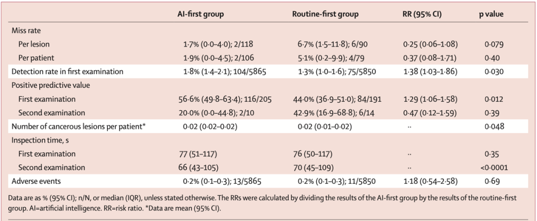
To evaluate whether this AI system can reduce the missed diagnosis rate of early esophageal squamous cell carcinoma and improve its detection rate in real clinical practice, Professor Hu Bing’s team initiated a multicenter, tandem, randomized controlled trial. This clinical trial involved 12 hospitals nationwide and 73 endoscopists. To ensure the quality of the research, all researchers participating in this trial underwent training related to the trial before it began, learning about the research protocol, the use of the AI system, the operation of the electronic random grouping table, and the data collection specifications for electronic case report forms. This study estimated the missed diagnosis rate and detection rate of early esophageal squamous cell carcinoma with and without AI system assistance based on previous research reports and pre-calculated the minimum sample size required for the trial (73 cases of early esophageal squamous cell carcinoma per group, totaling 146 cases; 4977 subjects per group, totaling 9954 subjects). From October 19, 2021, to June 8, 2022, a total of 11982 patients aged ≥18 years undergoing painless gastroscopy were continuously recruited. Participants were randomly divided into an experimental group (AI-assisted examination priority group, where AI assistance was available during the first esophageal examination and not during the second) and a control group (routine examination priority group, where AI assistance was not available during the first esophageal examination but was available during the second). A blind method was applied to subjects, pathologists, and data analysts, while no blinding was set for endoscopists, research assistants, and data collectors. The primary evaluation indicator was the missed diagnosis rate of early esophageal squamous cell carcinoma, while secondary evaluation indicators included the detection rate of early esophageal squamous cell carcinoma, positive predictive value, average number of early esophageal squamous cell carcinoma lesions per patient, examination time, AI false positive occurrence, and adverse event occurrence rate. A total of 11715 subjects were included in the final statistical analysis, with 5865 in the experimental group and 5850 in the control group. The study found that AI-assisted endoscopy achieved significant results in improving the detection rate of early esophageal squamous cell carcinoma, has the potential to reduce missed diagnoses, and demonstrated good safety (Figure 2, Table 1). This AI system is expected to become a powerful auxiliary tool in promoting the homogenization of diagnostic levels among endoscopists at all levels and improving the early diagnosis rate of esophageal squamous cell carcinoma.
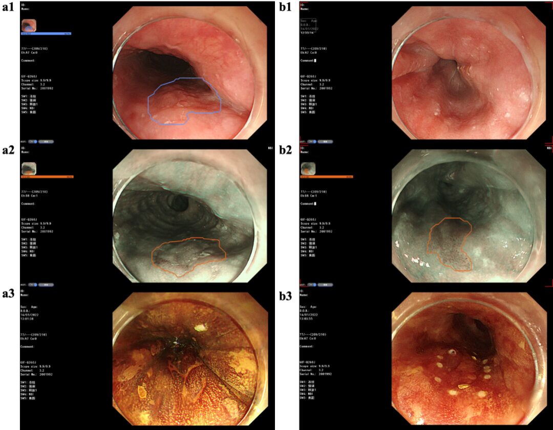
Figure 2: Example of Missed Lesions in the Routine Examination Priority Group (a) During the first examination without AI assistance, the doctor found one esophageal lesion invading the mucosal propria layer, which was subsequently detected by the AI-assisted doctor during the second esophageal examination under both white light mode and non-magnified narrow band imaging mode; (b) During the first examination without AI assistance, the doctor did not detect low-grade intraepithelial neoplasia, which was subsequently detected by the AI-assisted doctor during the second esophageal examination under non-magnified narrow band imaging mode. (a1, b1) Endoscopic images under white light mode; (a2, b2) Endoscopic images under non-magnified narrow band imaging mode; (a3, b3) Endoscopic images under iodine staining
Table 1: Comparison of Results Between Two Groups
Expert Commentary
Professor Alexander Arlt: Despite continuous innovations in gastrointestinal endoscopy equipment and widespread implementation of esophageal cancer screening projects, esophageal cancer remains one of the major malignant tumors threatening the lives and health of people worldwide, urgently requiring an improvement in its early diagnosis rate. With the cross-integration of gastrointestinal endoscopy and AI technology, AI-assisted diagnostic technology is expected to become one of the solutions for achieving early diagnosis and treatment of esophageal cancer. The highlight of this study is the development of an early esophageal cancer AI-assisted diagnostic system that integrates directly with the endoscope host and endoscope display, eliminating the need for an external display, and conducting the world’s first multicenter clinical trial to evaluate the real-time assistance of AI systems in diagnosing early esophageal cancer in a real clinical environment. The results of this study indicate that AI-assisted endoscopic examinations can significantly improve the detection rate of early esophageal cancer, suggesting that AI-assisted endoscopic diagnosis can effectively reduce the subjective bias of endoscopists’ visual diagnoses, achieving rapid and accurate diagnoses while providing an effective tool for training junior endoscopists.

Professor Alexander Arlt, Director of the Endoscopy Center at the University Hospital of Kiel, Germany, Member of the European Society of Gastrointestinal Endoscopy. His main research focuses on minimally invasive diagnosis and treatment of gastrointestinal diseases and pancreatobiliary diseases, having published dozens of papers in journals such as Nature Medicine, Nature Communication, GUT, Endoscopy. He studied advanced endoscopic techniques under Professor Hu Bing at West China Hospital, Sichuan University, in June 2013.
Author’s Insights
In recent years, with the rapid development of AI-assisted diagnostic technology, significant progress has been made in endoscopic diagnosis of early esophageal squamous cell carcinoma. Several studies have reported that AI can not only accurately detect early esophageal squamous cell carcinoma lesions but also correctly assess the extent and infiltration depth of lesions. Although past studies have shown good performance of these AI systems in diagnostic capabilities, they still have some limitations: (1) application is limited to a single scenario or function; (2) small or singular sample sizes that cannot fully simulate the heterogeneity of lesions; (3) primarily static image testing with limited real-time video testing; (4) lack of evaluation of AI systems’ auxiliary diagnostic performance in real, complex clinical environments. In response, our team has collaborated with universities and enterprises to focus on developing and optimizing AI-assisted endoscopic diagnosis models for early esophageal squamous cell carcinoma. After five years of research and development, we successfully transformed the laboratory AI model into a clinically applicable AI product. To evaluate the auxiliary diagnostic performance of this AI product, clinical trials in real environments are planned. Due to the inherently low detection rate of early esophageal squamous cell carcinoma in our country, a large minimum sample size is needed (73 cases of early esophageal squamous cell carcinoma per group, totaling 146 cases; 4977 subjects per group, totaling 9954 subjects). To quickly recruit enough subjects and assess the generalizability of AI in multicenter applications, a nationwide multicenter clinical trial was decided upon.
Given the large number of participating centers and daily patient recruitment, we have taken adequate measures for quality control in our research. First, all researchers participating in this trial underwent training related to the trial before it began, and we consulted statistical experts multiple times for valuable advice on selecting evaluation indicators. Then, during the implementation of the trial, research assistants contacted each sub-center daily to monitor research progress and, under the guidance of endoscopic experts, ensured data quality, holding regular progress meetings. After the trial concluded, we centralized and verified all collected data and completed data analysis under the guidance of statistical experts. Theoretically, AI is more effective in assisting junior doctors, but unfortunately, this study did not yield that result. We speculate that this may be due to not strictly limiting the skill level of operating doctors in the study design, resulting in a low number of junior doctors in both groups and a low number of lesions detected by junior doctors, with subgroup sample sizes insufficient to detect differences between the two groups, failing to reflect the true auxiliary effect of AI on junior doctors. Therefore, future large-sample clinical studies are still needed to specifically assess whether AI can improve junior doctors’ diagnostic levels for early esophageal squamous cell carcinoma.
The Lancet is one of the oldest and most influential medical journals in the world, and The Lancet Gastroenterology & Hepatology is a subsidiary journal of The Lancet, a leading journal in gastroenterology and hepatology, ranking 2nd among 93 journals in the JCR category of “Gastroenterology and Hepatology” with an impact factor of 35.7. This paper was submitted to The Lancet Gastroenterology & Hepatology in June 2023, and after initial review by the editor, it was recommended for publication via the fast track. The article was reviewed by four reviewers, including three medical experts and one statistical expert. The comments from the three medical experts mainly focused on issues regarding the inclusion and diagnostic standards of low-grade intraepithelial neoplasia, the selection of endoscopic modes, the sequence of iodine staining endoscopy, endoscopic quality control issues, and commercialization of the AI system. The statistical expert provided detailed suggestions on the choice of statistical methods to ensure the reliability and statistical rigor of the research results. After three rounds of revisions (each with a one-week deadline), the article was successfully accepted in August 2023 after carefully addressing the questions raised by the four reviewers and the editor. This journal has high standards for paper quality, requiring not only high-quality research content but also strict adherence to journal guidelines for writing abstracts and the main body structure, and it requires references to avoid citing articles published in various alert periodicals.
Corresponding Author
Hu Bing, PhD supervisor, Chief Physician of the Department of Gastroenterology at West China Hospital, Sichuan University. Standing member of the Gastrointestinal Endoscopy Branch of the Chinese Medical Association, Vice Chairman of the Pancreatic Disease Professional Committee of the Chinese Physician Association, Expert at the National Digestive Quality Control Center and Director of the Provincial Quality Control Center, Executive Director of the Sichuan Artificial Intelligence Society. Academic leader in Sichuan Province, national distinguished doctor, and famous doctor in Tianfu. Editor-in-Chief of the SCI journal World J of Gastrointest Endosc. His main research direction is early diagnosis and treatment of gastrointestinal tumors. As the chief scientist, he undertakes one national key research and development project and three general projects of the National Natural Science Foundation. In recent years, he has published over 160 SCI papers, ranking first in the number of SCI papers published in the field of gastroenterology and liver disease in the country for four consecutive years. He has over 30 authorized patents and successfully transformed 5, obtained 7 software copyrights, and 1 product registration certificate. As an internationally renowned endoscopy expert, he has been invited multiple times to demonstrate surgeries and teach in countries such as the United States, the United Kingdom, Germany, Canada, Russia, the UAE, India, and Egypt.
First Author
Yuan Xianglei, assistant researcher, postdoctoral fellow, physician at the Department of Gastroenterology, West China Hospital, Sichuan University. Member of the board of directors of the Sichuan Artificial Intelligence Society, standing committee member of the Gastroenterology and Gastrointestinal Endoscopy Collaborative Innovation Committee of the Sichuan Medical Innovation Association, and committee member of the Smart Endoscopy Branch of the Sichuan Bioinformatics Society. Mainly engaged in minimally invasive diagnosis and treatment of early gastrointestinal cancer and medical-engineering integration research. As the first/co-first author, he has published more than 20 SCI papers and participated in 8 authorized invention patents.
Co-First Author
Liu Wei, lecturer, postdoctoral fellow, physician at the Department of Gastroenterology, West China Hospital, Sichuan University. Vice Chairman of the Youth Committee of the Medical Robotics and Artificial Intelligence Branch of the Sichuan Physician Association, and member of the board of directors of the Sichuan Artificial Intelligence Society. His main research focuses on minimally invasive diagnosis and treatment of early gastrointestinal cancer and pancreatobiliary diseases, gastrointestinal endoscopy, and artificial intelligence. As the first/co-first author, he has published 20 articles in journals such as Lancet Gastroenterology&Hepatology, Endoscopy, obtained 7 authorized invention patents, and co-authored one book.
Co-First Author
Lin Yixiu, a resident in the Department of Internal Medicine at West China Hospital, Sichuan University, class of 2021. As the first/co-first author, she has published one paper in a Chinese core journal and one in an SCI journal, and participated in two invention patent applications.
Team Introduction
Professor Hu Bing’s team is a pioneer and leader in the field of early gastrointestinal cancer diagnosis and treatment both domestically and internationally, having conducted over 10 international first-in-class technology projects, establishing a database and biobank for early gastrointestinal cancer, and conducting interdisciplinary research across various fields such as endoscopic screening, early cancer diagnosis, preoperative assessment, ESD surgery, pathological features, postoperative follow-up, mechanisms of early cancer occurrence, epidemiological surveys, technological innovation, product development, and medical-engineering integration. The team has received 10 national projects and published multiple studies in the field of early cancer diagnosis and treatment, including basic research on esophageal organoids, endoscopic resection of giant esophageal leiomyoma, AI for early esophageal cancer, RCT of postoperative radiotherapy for early esophageal cancer after ESD, endoscopic 3D visualization technology, RCT for radiation protection during ERCP, AI for intestinal diseases, underwater capsule endoscopy, endoscopic appendectomy via the cecum, and magnetic bead-assisted traction technology. The developed product, the “AI-Assisted Diagnostic System for Early Esophageal Cancer,” was shortlisted for the Ministry of Industry and Information Technology’s innovation task for AI medical devices and was invited to participate in the national “13th Five-Year” science and technology innovation achievements tour exhibition, being selected as one of the first “Top Ten Advances in Medical Engineering at West China.” Team members have repeatedly won national early cancer competition awards, and their early cancer public education outreach is at the forefront in the country, recognized as a powerful team for early gastrointestinal cancer diagnosis and treatment both domestically and internationally.
Under the leadership of Professor Hu Bing, the gastrointestinal endoscopy center has developed into an internationally renowned training base for doctors from home and abroad, achieving multiple technological innovations and developing several innovative products. To date, dozens of physicians from countries such as the United States, the United Kingdom, Germany, Canada, Russia, the UAE, India, and Egypt have come to West China Hospital, Sichuan University, for training, including physicians from globally renowned hospitals such as Mayo Clinic, Johns Hopkins, Georgetown University Hospital, Southwestern Medical Center, Vanderbilt Medical Center, and the University of Kiel in Germany. Through technical exchange and cooperation with countries in Europe, America, and along the “Belt and Road” initiative, West China Endoscopy has taken the international stage, occupying the high ground of global endoscopic innovation technology.
Copyright Statement: The platform advocates respecting and protecting intellectual property rights. Reproduction and citation are welcome, but authorization from this platform is required. If you have any doubts about the copyright of the article content, please call 028-85422060, and we will communicate with you promptly. The content and images on this site are for reference and learning purposes only, not for profit and not to be used as diagnostic or medical basis.
Editor: Guo Qiong
Typesetting: Zhang Hongxue
Recommended Reading
1. The team led by Professor Hu Bing from the Department of Gastroenterology, West China Hospital, Sichuan University, and the team led by Professor Zhang Yi from the Intelligent Medicine Center of Sichuan University: AI Assistance Provides New Strategies for Quality Control in Colonoscopy
2. The team led by Professor Hu Bing from the Department of Gastroenterology, West China Hospital, Sichuan University: AI-Assisted Detection and Accurate Outlining of a Case of Micro Early Esophageal Cancer

Introduction to West China Journal
West China Journal was established in 2006 and publishes 14 medical technology journals (9 in Chinese and 5 in English), including 2 SCI-indexed journals, 2 ESCI-indexed journals, 1 EI-indexed journal, 4 MEDLINE-indexed journals, 7 CSCD-indexed journals, and 6 journals included in the overview of core Chinese journals by Peking University. West China Journal has won several awards including the “China Publishing Government Award Journal Award,” “China’s Top 100 Outstanding Academic Journals,” “China’s Boutique Science and Technology Journals,” and “China’s Most Internationally Influential Academic Journals.” One journal has been selected for the “China Science and Technology Journal Excellence Action Plan – Leading Journal” project, and two journals have been selected for the “China Science and Technology Journal International Influence Enhancement Plan – Class D (now the China Science and Technology Journal Excellence Action Plan – High Starting Point New Journal)” project. In the context of national double first-class construction, West China Journal is accelerating the construction of English journals and has established cooperative relationships with internationally renowned publishing groups such as Nature, Wiley-Blackwell, Oxford University Press, and BMC.
This article is reproduced from the WeChat public account West China Medical Time

Long press or scan the QR code to follow us!
Copyright Statement:This article was first published in the “Chinese Journal of General Surgery”. Other public accounts and dissemination media must contact the editorial department of this journal for authorization to reproduce, and mark “This article was published in the ‘Chinese Journal of General Surgery’, year, volume (issue): start and end pages or this article was published in the ‘Chinese Journal of General Surgery’, year, priority publication” in a prominent position. Thank you for your cooperation!
Click below “ Read the original text” to view other articles from our current issue