The Significance of GCC and GCA Examination
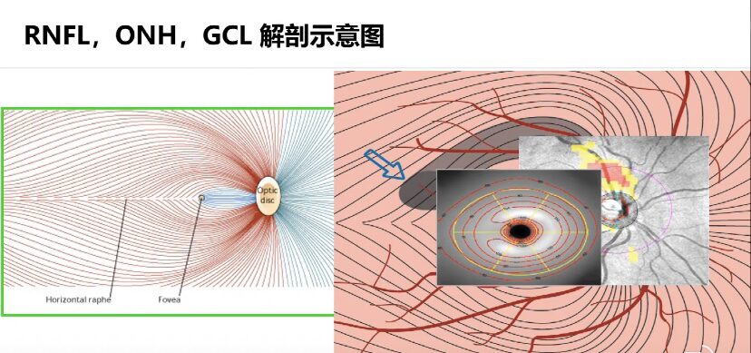
The structure parameters of the Circumpapillary retinal nerve fiber layer (cpRNFL) and the optic nerve have always been important detection indicators for OCT in the application of glaucoma diseases. However, due to the large range of optic nerve cup-to-disc ratio, changes in the vascular structure of the optic disc, and differences in the tilt angle of the optic disc, the reliability of cpRNFL measurement is still significantly affected.
With the development of Optical Coherence Tomography (OCT) technology, changes in the macular ganglion cell complex (mGCC) can be detected in an increasing number of ophthalmic diseases related to ganglion cell damage.
Since the structure of the macular region is relatively stable, and more than half of the retinal ganglion cells are located in the macular region, which are large and easy to detect, especially with the advancement of OCT technology, people have achieved more precise analysis of the inner structure of the macula, mainly the thickness of the Ganglion Cell Complex (GCC). Detecting mGCC has certain significance for early diagnosis of diseases, manifestations at various stages, treatment effects, and prognosis.

GCCRetinal Ganglion Cell Complex
Clinically, RNFL thickness and optic disc-related parameters are often used for diagnosing and assessing optic nerve damage. In addition to pRRNFL, retinal ganglion cells are also distributed in the ganglion cell layer and inner plexiform layer, which is the structure referred to as mGCC.
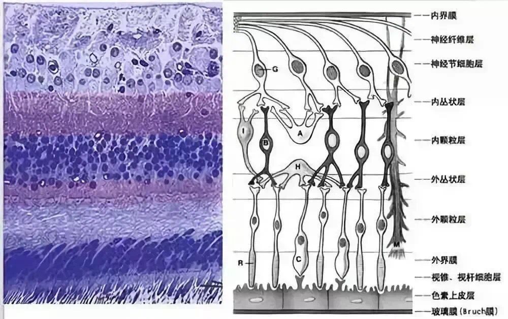
The macular region retinal ganglion cell complex (ganglion cell complex, GCC) includes the nerve fiber layer (RNFL), ganglion cell layer (GCL), and inner plexiform layer (IPL), representing the axons, cell bodies, and dendrites of RGCs, respectively. As glaucoma progresses, RGCs gradually decrease, leading to a thinning of GCC thickness. Its thickness and related analysis parameters can reflect the degree of damage to ganglion cells and exhibit different manifestations at various stages of different diseases.
Studies show that the cell bodies of retinal ganglion cells are distributed in only one layer in most areas, with 2-3 layers around the optic disc, and can have more than 8 layers in the macular region. As more than half of the retinal ganglion cell bodies are located near the macular region, the changes in mGCC thickness theoretically correspond more closely to the pathological structural changes of glaucoma than pRNFL thickness.

GCA Retinal Ganglion Cell Inner Plexiform Layer
GCA measurement is performed by removing the nerve fiber layer from the GCC and measuring the thickness of the ganglion cell + inner plexiform layer. Due to individual variability in the distribution of the nerve fiber layer in the macular region, it is difficult to determine whether a loss is due to a true defect or normal variation during statistical analysis. In comparison, the distribution of the ganglion cell and inner plexiform layer complex in the normal population is more regular, and this narrower range of variance will exhibit higher accuracy in statistical analysis. Therefore, based on the above research, GCA analysis that removes the nerve fiber layer can show higher statistical analysis accuracy.
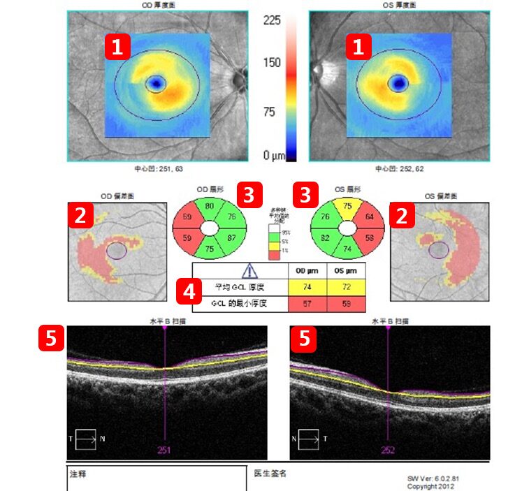
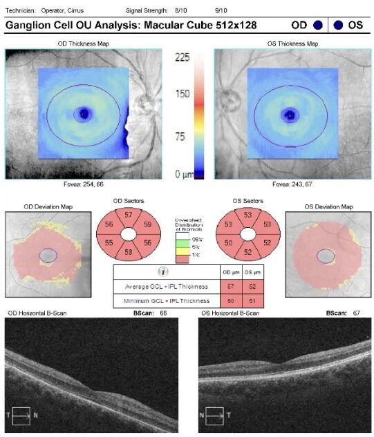
① Bilateral Thickness Map: Thickness color scale to show the thickness of the ganglion cell + inner plexiform layer in the elliptical area of the macular region
② Bilateral Thickness Deviation Map: Red-yellow areas indicate regions where the ganglion cell complex is thinned.
③ Six-Quadrant Thickness Values: Green indicates this quadrant value is within the normal range, yellow indicates borderline, red indicates thin, and white indicates thick.
④ Data Table: Average thickness values and minimum thickness values of the ganglion cell complex in the bilateral macular region.
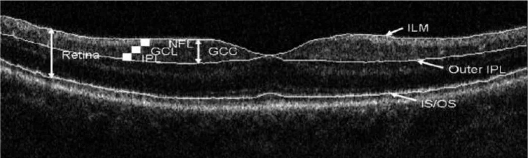
⑤ Bilateral Macular Ganglion Cell Complex Tomography: Boundary positions (purple line and yellow line) for measuring the thickness of the ganglion cell complex.

Thank you for watching
Long Press the QR Code
Follow Us

WeChat ID: Eye Specialty Examination Learning Network
Gain a little every day