

The gamma camera is a photographic system that records and displays the distribution of radiation activity within an imaging object. It collects gamma rays emitted from the patient’s body and reconstructs an image of the emission site, aiding in the understanding of the functions of specific organs or systems in the human body. Unlike a scanner, it does not have mechanical scanning parts, and the time required for imaging is very short; capturing a frame takes only a fraction of a second, and it can automatically take continuous photographs, showing dynamic changes in the distribution of radionuclides.


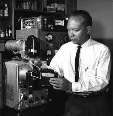
The gamma camera was designed by Hal O-Anger in the late 1950s and is therefore also known as the Anger camera. The gamma camera has a large imaging field and can record both static and dynamic images of patients, offering capabilities that are irreplaceable by other diagnostic instruments. After the 1970s, the development of the gamma camera accelerated rapidly, and together with CT and ultrasound, it formed a new discipline—medical imaging. By 1982, compared to ten years prior, research in nuclear medicine on the heart, liver, and bones had increased, while research on the lungs remained at previous levels, and studies on organs such as the brain and kidneys had decreased, which accelerated the application of the gamma camera in clinical settings.


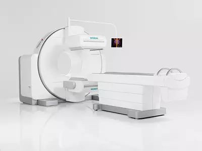
The gamma camera consists of a collimator, sodium iodide (thallium) crystal, light guide, photomultiplier tube matrix, positioning circuit, energy circuit, display system, and imaging device. The collimator, crystal, and photomultiplier tube matrix form the movable parts known as the probe, which is the core of the gamma camera. During operation, the gamma rays emitted from the radioactive nuclide injected into the body first pass through the collimator, then strike the sodium iodide crystal, generating flashes of light that are collected by a group of photomultiplier tubes. The corresponding weighting and integration of all photomultiplier tubes can produce positional and energy signals. The processed signals become a count record, forming an image of the radioactive concentration distribution in the body, which is a gamma camera image.


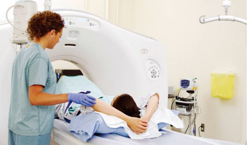
The gamma camera is the most basic and important imaging device in modern nuclear medicine. It performs a single imaging of the distribution of radioactive nuclides in internal organs and allows for dynamic observation. The gamma camera can track and record the morphology and function of radioactive drugs passing through specific organs through continuous imaging, making it suitable for dynamic studies; due to the relatively short examination time, it is convenient and simple, especially suitable for children and critically ill patients; imaging is rapid, facilitating multi-position and multi-site observations; by processing the images accordingly, it can provide diagnostic data or parameters. Clinically, it can be used for static dynamic imaging examinations of organs, with dynamic imaging primarily used for cardiovascular disease diagnostics.


Before the advent of SPECT and PET, the gamma camera was a highly advanced nuclear medicine imaging instrument. As a non-invasive diagnostic tool, it plays an important role in the clinical diagnosis of diseases of organ systems such as the cardiovascular system, liver, orthopedics, kidneys, and brain.
Cardiovascular Diseases
Cardiovascular diseases are the main application area of the gamma camera, which is divided into two major categories: radioactive cardiovascular angiography and myocardial imaging. The former can determine ventricular function, including important cardiac parameters such as ventricular ejection fraction, wall motion, and peak filling rate during diastole, and can study the dynamic changes of radionuclides as they pass through the heart’s chambers. The latter utilizes the selective absorption of myocardial equivalent T1 tracers (cold spot imaging) or the selective accumulation of tracers such as Tc pyrophosphate in damaged myocardium to estimate cardiac conditions such as myocardial ischemia, myocardial infarction, left ventricular wall aneurysm, and congenital heart disease.
Liver and Gallbladder Diseases
The gamma camera was once the preferred method for liver imaging because it could easily display the entire morphology of the liver, facilitating clinical observation, and had a lower false-negative rate. Especially when the patient has “artifacts” due to respiratory motion, both CT and ultrasound examinations are limited, and only imaging can make a diagnosis. In liver cancer patients, it is common for the density difference between cancer tissue and normal liver cells to be small; in this case, CT can easily misdiagnose, while the gamma camera can often clearly indicate the presence of a tumor. However, the gamma camera may not easily detect benign or malignant space-occupying lesions smaller than 2 cm. Therefore, it is generally believed that accurate diagnosis of liver diseases should combine gamma imaging, ultrasound, and CT methods.
Cerebrovascular Diseases
For the diagnosis rate of intracranial space-occupying lesions, overall, CT is slightly higher than that of gamma camera imaging, but the degree of difference varies among different diseases. Statistics from abroad on 238 cases of intracranial space-occupying lesions indicate that the diagnostic rate for CT is 92.8%, while for the gamma camera it is 85.3%. However, in groups such as meningiomas, metastatic tumors, and abscesses, the diagnostic rates of both are essentially the same. In groups such as subdural hematomas and arteriovenous malformations, the diagnostic rate of gamma camera imaging is even higher than that of CT. However, gamma camera imaging is superior to CT in reflecting the metabolic status of blood flow, as the imaging of cerebral thrombosis can detect all cases, whereas the positive rates for EEG and cerebral blood flow imaging are 60% each.
Kidney Diseases
Dynamic renal imaging is particularly valuable for understanding renal function, such as urinary tract obstruction, where gamma camera imaging can provide a comprehensive and accurate diagnosis; monitoring of transplanted kidneys can also be timely and accurately reflected by gamma camera imaging.
With the advancement of medical technology, in order to achieve more accurate diagnoses, the gamma camera has gradually been phased out, with SPECT, PET, and PET/CT imaging examinations being able to reflect normal or abnormal physiological metabolic activities in the body more accurately. SPECT is a tomographic imaging method based on gamma camera technology, still falling under the gamma camera category. It involves one or more probes rotating 360 degrees around a specific organ of the patient, capturing a frame of images at regular intervals, and then overlaying and reconstructing the images into cross-sections or any required orientations, greatly enhancing the sensitivity and accuracy of diagnoses. (Source: “Guangdong Radiation Protection” December 2016, Issue No. 18)

Guangdong Radiation Protection Association Your Radiation Consultant!
WeChat ID: gdsfsfhxh

(Long press to scan the QR code and easily follow us!)
Welcome to comment, forward, and share, so that together with those around you, you can understand radiation better and protect yourself!